EMBRYOLOGY
The endodermal diverticulum off the foregut grows into the septum transversum; the paired vitelline veins also grow into the septum transversum, divide into a vascular network and then reunite as a pair of veins, into the sinus venosus.
Parenchymal cells arise from the endoderm.
Sinusoids arise from vitelline and umbilical venous elments.
Connective tissue elements and fibrous capsule arise from septum transversum.
Bile ducts and the gallbladder arise from endodermal diverticulum.
Fetus:
The liver is not fully active–the left umbilical vein uses the ductus venosus to bypass the liver to enter into the ivc. The left and right lobe are almost equal in size.
childhood
adulthood
old age
Introduction
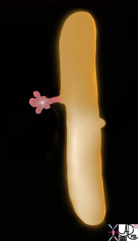
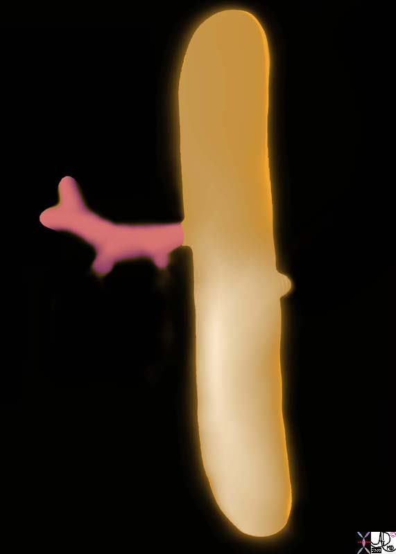
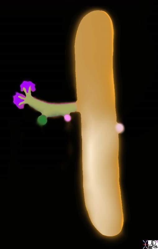
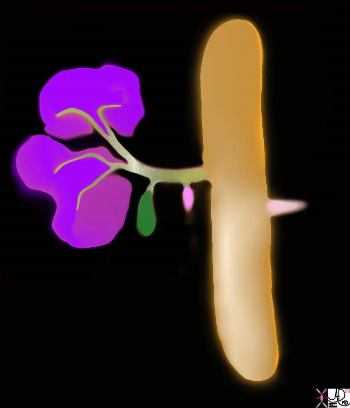
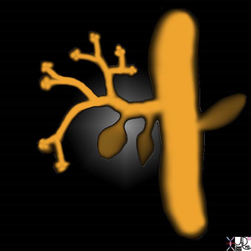
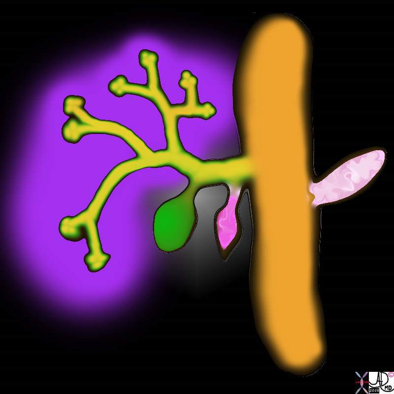
In the 4th week of gestation, the primitive endoderm gives rise to a foregut diverticulum called the hepatic diveticulum at the junction of the of the foregut and midgut. The hepatic diverticulum is the precursor for the liver, bile ducts. and gallbladder. The hepatic diverticulum gives rise to a pars hepatica and pars cystica. The pars hepatica will evolve into the liver and bile ducts while the pars cystica will develop into the gallbladder and cystic duct. The endodermal bud is initially a solid cord but by week 8 with growth and resorbtion the tubular components are formed. These tubular components grow into the mesodermal component of the liver called the septum transversum.
|
Hepatic Diverticulum |
|
In the 4th week of gestation, the primitive endoderm gives rise to a foregut diverticulum called the hepatic diveticulum at the junction of the of the foregut and midgut. The hepatic diverticulum is the precursor for the liver, bile ducts, and gallbladder. liver bile ducts pars cystica gallbladder and cystic duct. endodermal bud solid cord growth and resorbtion gallbladder embryology normal Davidoff art copyright 2008 82219a01b2.8s 82219a02a1.8s |
|
Hepatic Diverticulum |
|
Aftter the 4th week of gestation, the primitive endoderm continues to differentiate and grow. branches gives rise to a foregut diverticulum called the hepatic diveticulum at the junction of the of the foregut and midgut. The hepatic diverticulum is the precursor for the liver bile ducts and gallbladder. hepatic diverticulum pars hepatica and pars cystica. liver bile ducts pars cystica gallbladder and cystic duct. endodermal bud solid cord growth and resorbtion gallbladder embryology normal Davidoff art copyright 2008 82219a02b1.8s 82219a02b3.8s |
complex interactions between the mesoderm and the epithelial cells of the endoderm take place. It is only with the formation of the vessel system and the development of the portal vein that the definitive organ structure is assembled.
|
Hepatic Diverticulum |
| In the 4th week of gestation, the primitive endoderm gives rise to a foregut diverticulum called the hepatic diveticulum at the junction of the of the foregut and midgut. The hepatic diverticulum is the precursor for the liver bile ducts and gallbladder. hepatic diverticulum pars hepatica and pars cystica.
liver bile ducts pars cystica gallbladder and cystic duct. endodermal bud solid cord growth and resorbtion gallbladder embryology normal Davidoff art copyright 2008 82219b01.8s |
|
Hepatic Diverticulum |
| In the 4th week of gestation, the primitive endoderm gives rise to a foregut diverticulum called the hepatic diveticulum at the junction of the of the foregut and midgut. The hepatic diverticulum is the precursor for the liver bile ducts and gallbladder. hepatic diverticulum pars hepatica and pars cystica.
liver bile ducts pars cystica gallbladder and cystic duct. endodermal bud solid cord growth and resorbtion gallbladder embryology normal Davidoff art copyright 2008 82219b08.8s |
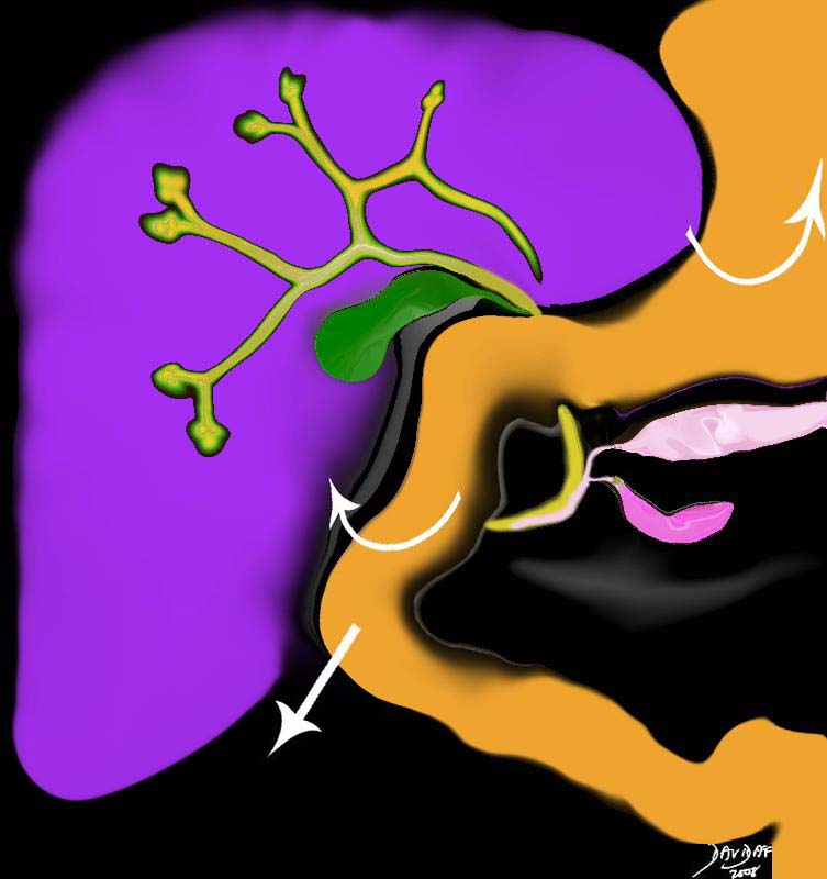
As the stomach grows it rotates to the left and the the duodenum grows down and to the right and also rotates to the right. The combination of these three events brings the distal common bile duct, and ventral pancreas to the medial and left side of the duodenum.
The CBD and Ampulla on the Medial Duodenum |
| In the 5th week, the duodenum has grown down and to the right, as well as rotated 90 degrees to the right. The stomach rotates to the left. These events bring the pancreatic duct and the CBD to the posterior and medial aspect of the duodenum.
82219b12.8s liver gallbladder pancreas foregut midgut bile duct cystic duct common bile duct stomach embryology septum transversum mesoderm endoderm 5th week stomach rotated to left duodenum growth |
o the left
Duplicated Gallbladder |
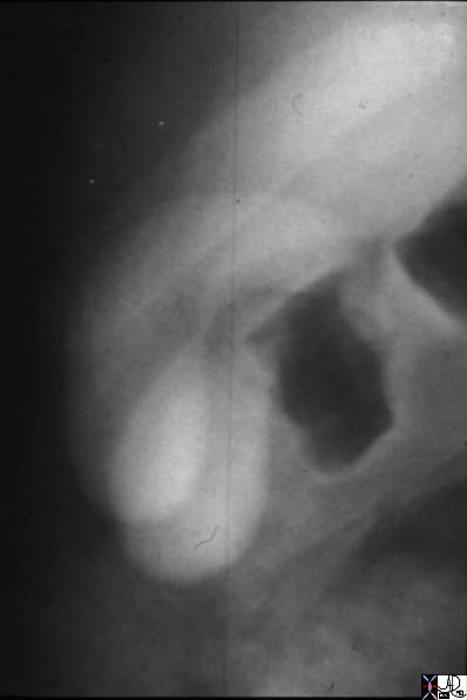
| 04750 gallbladder duplicated gallbladders oral cholecystogram congenital duplication anomaly growth Davidoff MD |
References
Meilstrup JW Imaging Atlas of the Normal Gallbladder and Its Variants By Jon W. Meilstrup Published by CRC Press, 1994
The development of the liver
Originally an exocrine organ reorganized its structural plan to perfect activities that are primarily related to the blood stream. (Arey) embryology endodermal diverticulum off the foregut grows into the septum transversum the paired vitelline veins also grow into the septum transversum, divide into a vascular network and then reunite as a pair of veins, into the sinus venosus parenchymal cells arise from the endoderm sinusoids arise from vitelline and umbilical venous elments connective tissue elements and fibrous capsule arise from septum transversum bile ducts and gallbladder arise from endodermal diverticulum fetus liver is not fully active the left umbilical vein uses the ductus venosus to bypass the liver to enter into the ivc left and right lobe are almost equal in size childhood adulthood old age regeneration Hepatic cells are replaced rarely in the normal, adult liver. marked capacity for repairing even extensive tissue losses. losses from toxic agents or surgery are recouped (as to liver weight) promptly. new lobules bud out of old ones, hepatic cells enlarging and increasing by mitosis. Bile ducts also proliferate and probably also form some new hepatic cells.
| Embryplogy |
| (Image courtesy of Ashley Davidoff M.D.) |

