Copyright 2012
Definition
Brain
AVM in the Posterior Fossa |
|
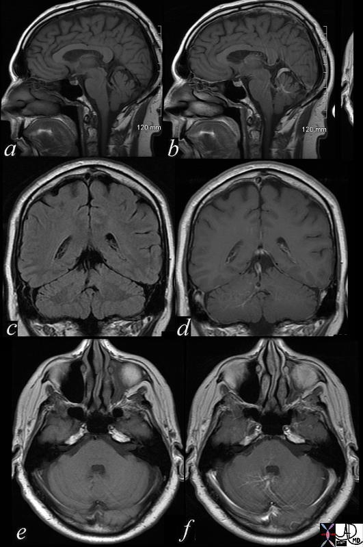
brain cerebellum vermis vein enlarged venous angioma old bleed superior left vermis – cerebellar venous malformation vascular malformation a = T1 pre gadolinium b = T1 post contrast c = T1 pre gadolinium d = T1 post contrast e = T1 pre gadolinium f = T1 post contrast MRI Copyright 2012 Courtesy Ashley Davidoff MD 71730c02 |
Hemorrhage in an AVM |
|
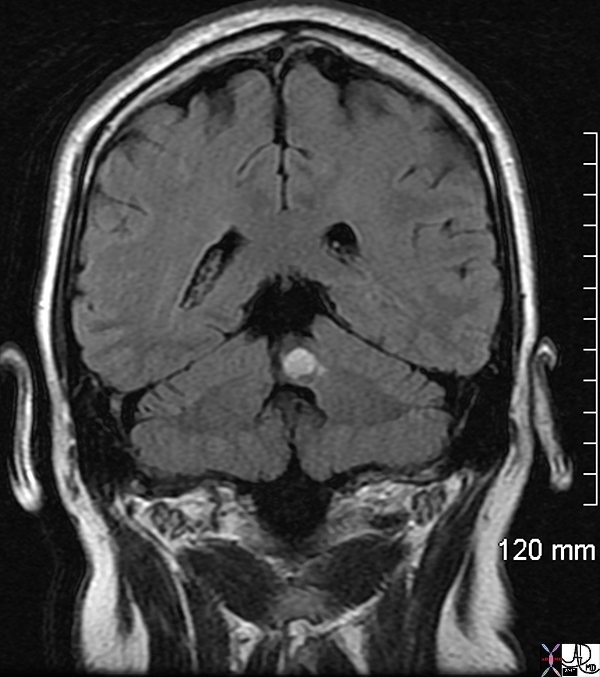
male with acute ataxia roof of fourth 4th ventricle acute bleed left vermis venous angioma brain cerebellum vermis vein cerebellar venous malformation vascular malformation T1 pre gadolinium T1 bright lesion dx acute hemorrhage MRI Copyright 2012 Courtesy Ashley Davidoff MD 71730 |
CVS
Patent Ductus Arteriosus |
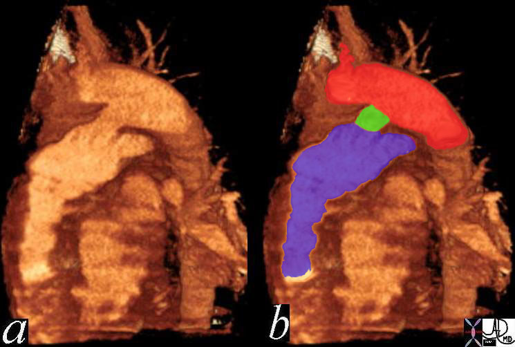
| Unsuspected shunt in a young adult male heart right ventricle RV and pulmonary artery = blue PDA = green Aorta = red patent ductus arteriosus CTA CTscan
Copyright 2012 Courtesy Ashley Davidoff MD 42326c01 |
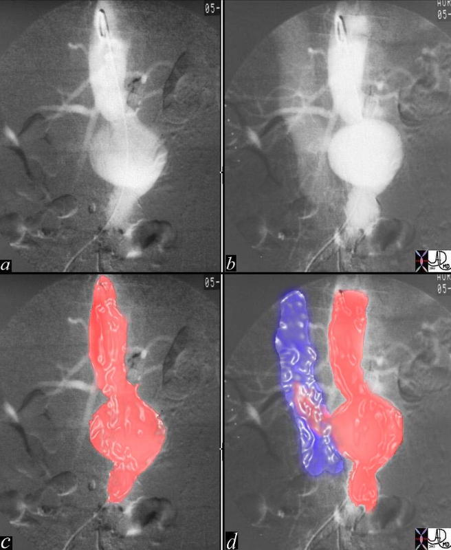
Arteriovenous Fistula Between the Aorta and Inferior Vena Cava |
|
This series of digital aortograms shows a rotund fusiform infrarenal aortic aneurysm (a, c – red overlay)) which later in the study shows premaure filling of the IVC (b, d blue overlay). This is an example of aneurysmal erosure of the the IVC and secondary formation of an arteriovenous fistula. Copyright 2012 Courtesy Laura Feldman MD 35858c03CP |
Adrenal

Arteriovenous Shunting in a Pheochromocytoma |
| The aortogram of the right kidney in the arterial phase (a,b) and nephrogram phase (c,d) reveals a hypervascular mass arising from the right adrenal gland overlaid in green (b and d). The salmon pink arrow in b shows the renal artery in the arterial phase and the diffuse homogeneous enhancement in the parenchymal or nephrogram phase is shown by the maroon arrow in d. The suprarenal mass shows different perfusional characteristics that typify arteriovenous shunting in the mass. In the arterial phase (a,b) the arterial supply (bright red) shows bizarre angulation, non uniform branching with early draining veins (blue) seen while the kidney is still in the arterial phase. The veins become more prominent while the renal vein of the kidney is yet to materialize. The mass was shown pathologically to be a pheochromocytoma. Angiography is not safely performed in patients with pheochromocytoma since a crisis can be precipitated by the contrast. This case originates from an era prior to sophisticated imaging and biochemical evaluation.
Copyright 2012 Courtesy Ashley Davidoff MD 26986cL02.9 |