The liver is pyramidal in shape with its base at the right and its apex toward the left.
The superior surface is smooth, and conforms to the dome-shaped contours of the hemidiaphragm.
The inferior surface tends to be flat, but there are undulations caused by moulding of the liver to the abdominal organs which relate to its inferior surface.
The right lobe is quadrilateral in shape while the left lobe is tongue-shaped with a relatively thin inferior margin.
Capsular depressions due to uneven development of the lobes or due to the impression of the diaphragmatic insertions may occur.
Riedel’s lobe is an inferior extension of the right lobe of the liver, occurring more commonly in women.
It is a normal anatomic variant and its presence changes both the size and shape of the right lobe of the liver.
Accessory lobes of the liver may occur anywhere in the liver but most occur off the right lobe of the liver.
Shape
The liver is pyramidal in shape with its base at the left and its apex toward the right. The external surface of the liver is smooth with the apex rounded over, similar to a large mushroom cap. (see diagram below)
|
Normal liver – falciform ligament |
|
Image courtesy of LifeArt Lippincott, Williams & Wilkins All rights reserved The pyramidal and mushroom shape of the liver is exemplified by this drawing. The falciform ligament runs an oblique course between the medial and lateral segments of the liver. At its inferior edge is a rounded ligament called the ligamentum teres. It is the remnant of the umbilical vein that brought maternal and placental blood to the developing fetus and baby. |

In cross sectional imaging, the surface of the liver is interrupted by normal notches and grooves. The most obvious are the grooves and fissures within the porta hepatis and the region of the entry point of the falciform ligament.
|
Notches |
|
The anterior smooth surface of the liver is interrupted by the notch caused by the entrance of the falciform ligament. Opposite this notch is a corresponding “internal” notch. These notches separate the medial (IV) and lateral (II & III) segments of the left lobe. Other internal notches are the interlobar groove which separates the right and left lobes, (IV & V) and the notch which reflects the position of the right hepatic vein. This notch indicates the line of division between the anterior and posterior segments of the right lobe.(in this case V & VI) (Image courtesy of Ashley Davidoff M.D.) |

In the following image, the thick blue line runs to the interlobar fissure and corresponds to the border between the right and left lobes as well as the expected location of the middle hepatic vein and gallbladder axis. The thin blue line runs between the border of the anterior and posterior segments of the right lobe and corresponds to the expected location of the right hepatic vein.
|
Obliterated umbilical vein |
|
Of special note: The yellow dot in the falciform ligament represents the obliterated umbilical vein which is a remnant of the embryologic left umbilical vein which transported blood from the mother to the developing fetus. The thick blue vein runs to the interlobar fissure and corresponds to the border between the right and left lobes, as well as the expected location of the middle hepatic vein and gallbladder axis. The thin blue line runs between the border of the anterior and posterior segments of the right lobe and corresponds to the expected location of the right hepatic vein. (Image courtesy of Ashley Davidoff M.D.) |

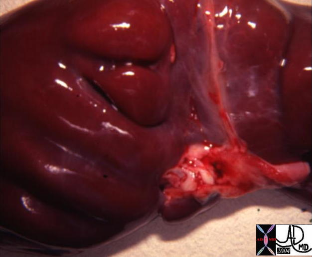
We reviewed the following image when we discussed the malleability of the liver, indicating that the tendons of the diaphragm were able to indent the soft liver.
|
Lobulated liver |
|
In this instance we se the example to show a variation of the normal smooth, and mushroom shaped surface. This lobular contour is a normal variant. |

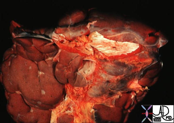
Compare the previous normal variant with the abnormal lobular contour of this liver. There is a distinct lack of uniformity with coarse micronodular and macronodular change. This is an example of severe cirrhosis.
|
Lobulated liver |
|
This liver has suffered the consequences of cirrhosis, in which the liver tissue is replaced by scar tissue resulting in an overall shrinkage of the liver. |

This image shows only micronodular change at the surface of the liver.
|
Congenital Fetal Lobultion |
| 13446 liver hepatic applied biology shape lobulation anatomy Davidoff MD |
| Hepar Lobatum |
| 13454 liver applied biology shape anatomy hepar lobatum congenital syphilis infection fibrosis Davidoff MD Courtesy Barbara Banner MD |
|
Lobulated liver |
|
This image highlights the abnormal area. Compare this image to the previous normal images to review the normal vs. abnormal contour of the liver. (Image courtesy of Ashley Davidoff M.D.) |
.jpg)
The highlighted area in the image below reveals micronodular cirrhosis, a characteristic of alcoholic cirrhosis.
|
Lobulated liver |
|
This image highlights the abnormal area. Compare this image to the previous normal images to review the normal vs. abnormal contour of the liver. (Image courtesy of Ashley Davidoff M.D.) |

Position
The liver resides in the upper and right quadrants of the abdominal cavity, nearly occupying the entire right hypochondrium, the greater part of the epigastrium, and not uncommonly extending into the left hypochondrium as far as the mammillary line.
|
Position of the liver |
|
This plain film of the abdomen shows the position of the liver in the abdominal cavity. Note it lies just below the right hemidiaphragm and it occupies almost the entire right upper quadrant. The metallic object in the center of the abdomen is an umbilical ring. (Image courtesy of Ashley Davidoff M.D.) |

|
Position of the liver |
|
The thick blue line that traverses the green gallbladder represents the position of the middle hepatic vein that divides the liver into right and left lobes. The thinner line represents the position of the falciform ligament which divides the left lobe into a medial rightward segment (IV)and a lateral leftward segment. (II and III) Part of the left lobe usually lies toward the right side. (Image courtesy of Ashley Davidoff M.D.) |
.jpg)
The left lobe is usually mostly on the right side of the body. Sometimes the lateral segments of the left lobe will cross the midline to lie leftward of the midline. The medial segment, segment IV, usually remains on the right side.
|
Position of the liver |
|
In this cross sectional image the liver is seen in relation to the J shaped stomach just medial the spleen posteriorly and the colon laterally. It occupies almost the entire right upper quadrant. (Image courtesy of Ashley Davidoff M.D.) |

Although unusual, some day you may come across a patient with situs inversus, which can make you look twice. In these patients, the position of the organs are reversed, with the liver on the left!
|
Position of the liver |
|
This image shows a contrast enhanced CT of a patient with situs inversus where the position of the internal organs are reversed from right to left and left to right. Notice the liver on the right side of the image and the left side of the body. (Image courtesy of Ashley Davidoff M.D.) |
.jpg)
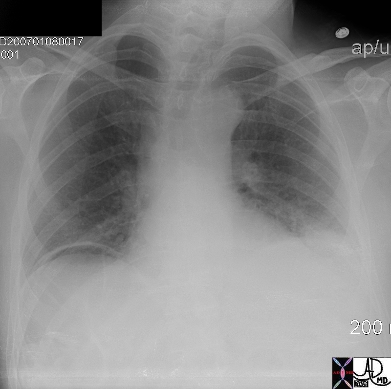
|
Question free air? |
|
This image shows a curvilinear air shadow that suggests free ar under the diaphragm. The patient was asymptomatic and so a subsequent upright KUB was performed. See next image (Image courtesy of Ashley Davidoff M.D.) 45758 45759 |
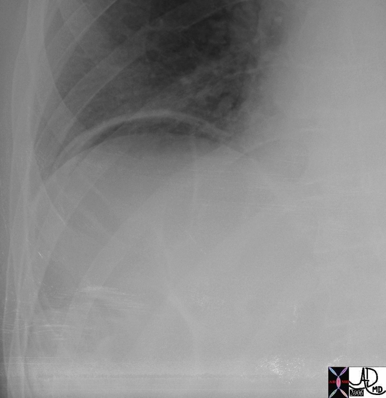
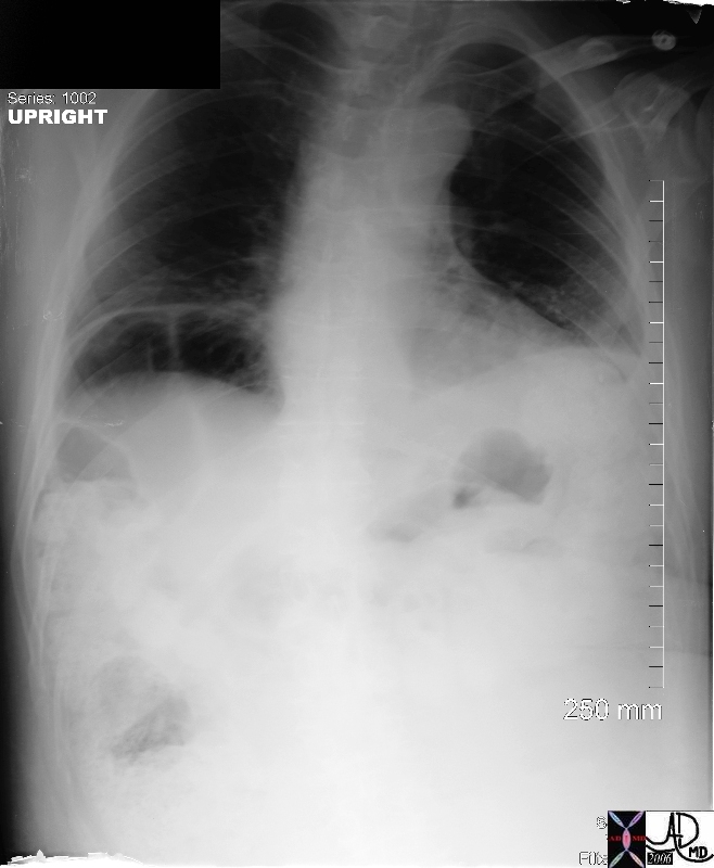
|
Question free air? |
|
This upright iamge of the abdomen clearly shows that the air under the diapragm is part of air within the lumen of a loop of bowel in the right upper quadrant. (Image courtesy of Ashley Davidoff M.D.) 45761 |
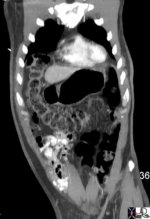
|
Chilaiditi syndrome – interposition the colon |
|
This coronal image of the abdomen shows the colon and specifically the hepatic flexure malpositioned under the diaphragm and mimics the presence of free air under the diaphragm as noted in the above images. The liver is displaced from its usual position by the colon which makes its way anteriorly (in front of the liver), and superiorly (below the diaphragm), so that if there is air within this loop it will masquerades as “free air” (Image courtesy of Ashley Davidoff M.D.) 45765 |
|
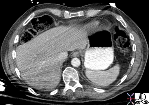
Chilaiditi syndrome – interposition the colon |
|
This axial image of the abdomen shows the colon and specifically the hepatic flexure malpositioned anterior to the liver. The liver is displaced from its usual position by the colon which makes its way anteriorly (in front of the liver), and superiorly (below the diaphragm), so that if there is air within this loop it will masquearades as “free air” (Image courtesy of Ashley Davidoff M.D.) 45768 |