Copyright 2012
Introduction
Atherosclerosis
Atherosclerosis and Thrombosis in the Coronary Artery |
|
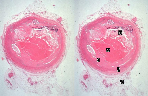
heart cardiac coronary artery fx narrowing fx stenosis fx plaque fx eccentric plaque a= thrombus b= cholesterol plaque c= fibrous plaque d= media e = adventitia dx atherosclerosis thrombosis histopathology Courtesy Dr Joris 13318c |
Atherosclerosis Stenosis |
|
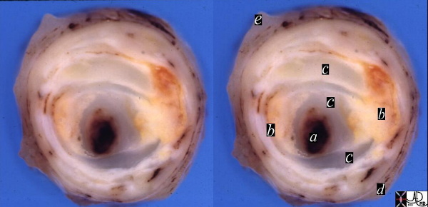
artery + coronary artery fibrocalcific plaque calcification narrowing stenosis dx atherosclerosis + grosspathology Courtesy Henri Cuenoud MD a= lumen or thrombus dx atherosclerosis + grosspathology Courtesy Henri Cuenoud MD 13410c |
Stenosis of the Left Renal Artery |
|
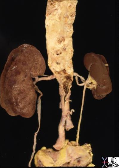
aorta abdomen iliac artery fx atherosclerosis kidney fx small dx RAS renal artery stenosis grosspathology Copyright 2012 Courtesy Ashley Davidoff MD 13417 |
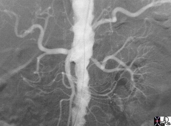
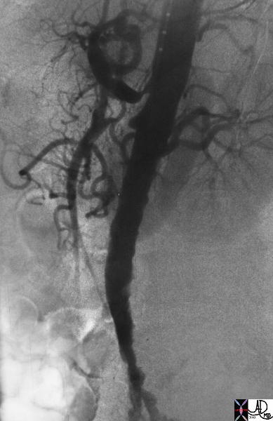
High Grade Stenosis of the Left Renal Artery |
|
aorta abdominal aorta atherosc;erosis atheroma renal artery stenosis kidney artery renal artery occlusion angiogram angiography aortogram aortography Copyright 2012 Courtesy Ashley Davidoff MD 16694.b01 |
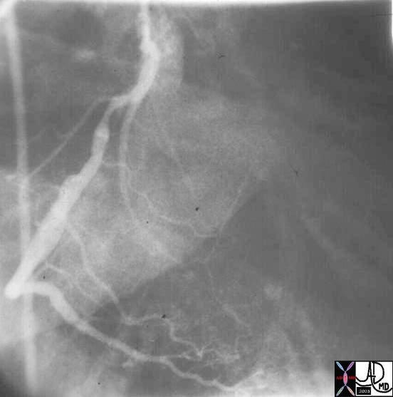
Stenosis of the Right Coronary Artery |
|
cardiac heart coronary artery RCA RAO PDA moderate stenosis angiogram angiography Copyright 2012 Courtesy Ashley Davidoff MD 15024 |
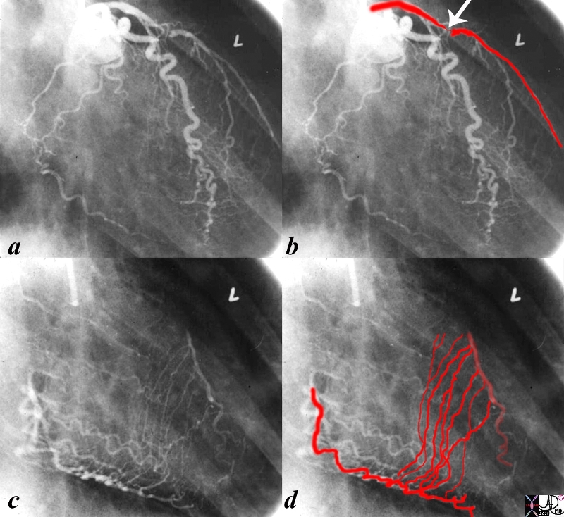
High Grade Stenosis of the LAD and Collateral Supply |
|
The first angiogram is from a an injection of the left coronary artery in the RAO projection. A subtotal stenosis is seen in the proximal LAD (arrow). There is an implied high pressure gradient between the proximal and distal LAD and a predicted low pressure in the distal vessel. The second injection was into a normal right coronary artery with delayed iaging when most of the contrast had disappeared from the vessel and shows septal collateral vessels supplying the downstrem low pressure LAD from a high pressure RCA. code heart cardiac stenosis atherosclerosis collaterals septal collaterals coronary artery septal perforators angiogram angiography Courtesy Ashley Davidoff MD copyright 2009 all rights reserved 15042c02.8s |
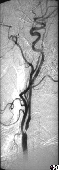
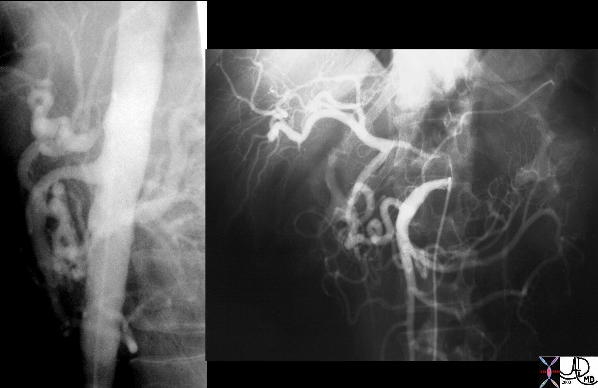
High Grade Stenosis of the Internal Carotid Artery |
| The selective angiogram of the the common carotid shows a high grade subtotal occlusion of the origin of the internal carotid artery. This is likely due to atherosclerosis. artery brain internal carotid fx stenosis subtotal occlusion at origin dx atherosclerosis atheroma angiogram angiography
Copyright 2012 Courtesy Ashley Davidoff MD 10218 |
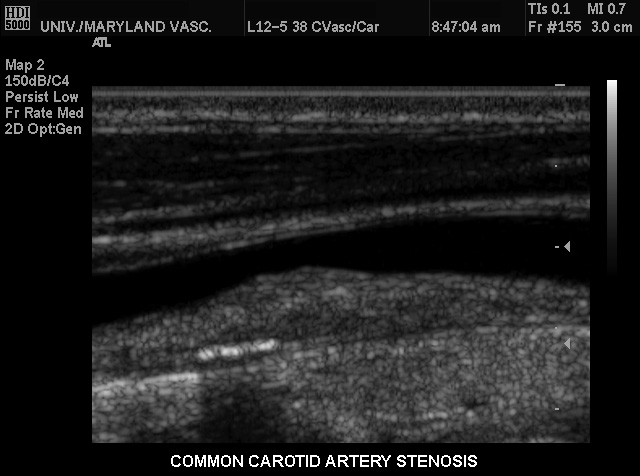
Ultrasound of the Common Carotid Revealing Stenosis |
|
This conventional gray scale US of the neck shows a common carotid artery heaped up plaque on the far wall causing stenosis of moderate degree. Note also the linear area of strong echogenicity on the far wall associated with shadowing consistent with a calcification in the wall. These findings are characteristic of atherosclerotic plaque. Courtesy Philips Medical Systems 33289 |
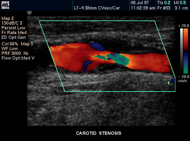
Color Flow Doppler of a Common Carotid Stenosis |
|
Patient with carotid artery stenosis demonstrated by color flow doppler. Courtesy Philips Medical Systems 33278 |
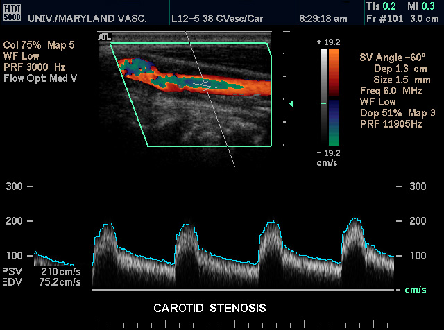
Atherosclerosis Stenosis – Pulse Interrogation |
|
This color flow doppler US with pulse flow interrogation of the carotid artery in the neck demonstrates small smooth focal bulges on either side of the lumen causing mild stenosis. This finding is characteristic of atherosclerosis. Courtesy Philips Medical Systems 33290 |
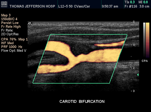
Power Flow Doppler Common Carotid Stenosis |
|
Patient with a stenosis of the distal common carotid artery at the level of the bifurcation demonstrated by color power doppler US technique . Courtesy Philips Medical Systems 33259 |
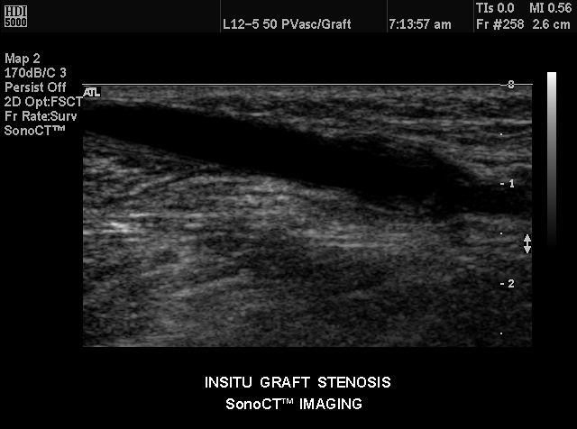
Graft Stenosis |
|
In situ graft stenosis of an artery by compound US. Courtesy Philips Medical Systems 33237 33242 |
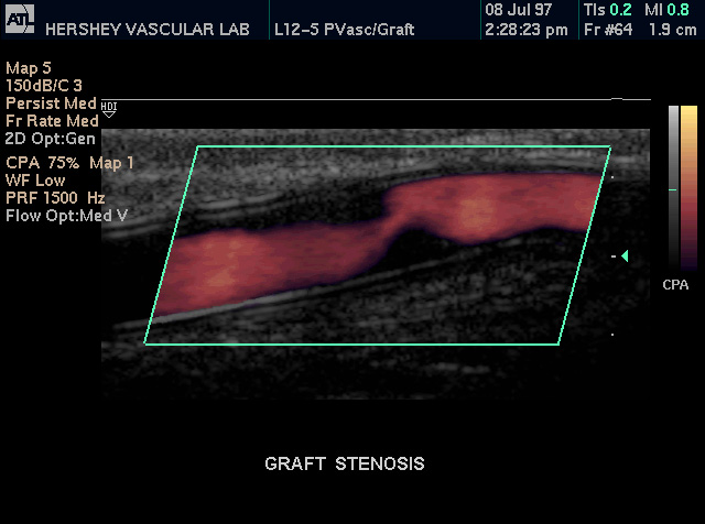
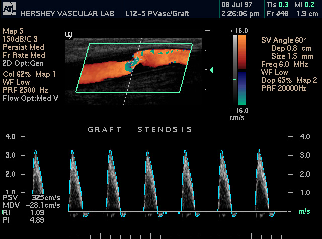
Doppler – High Grade Stenosis |
|
Graft stenosis shown by color doppler and high velocity pulse doppler US waves are shown at the stenotic site. Courtesy Philips Medical Systems 33243 |
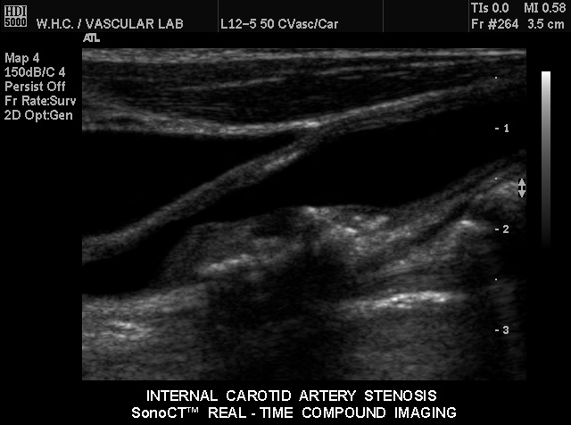
Calcified Atherosclerotic Plaque |
|
This gray scale US of the neck shows an area of thickening and heaping up off the far wall of the of the internal carotid artery characteristic of atherosclerotic plaque. There is a moderate stenosis of the vessel. Courtesy Philips Medical Systems. 33299 |
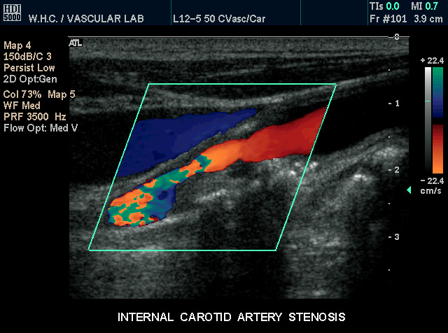
Color Flow Doppler of Stenotic Plaque |
| This color flow doppler US of the neck shows an area of thickening and heaping up off the near wall of the of the internal carotid artery characteristic of atherosclerotic plaque. There is a moderately severe stenosis of the vessel.
Courtesy Philips Medical Systems. 33302 |
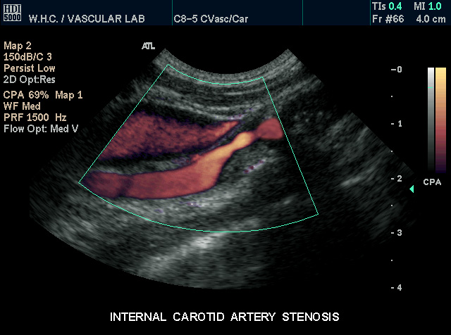
Power Doppler of Internal carotid Atherosclerotic Stenosis |
|
This power flow doppler US of the neck shows an area of thickening and heaping up off the near wall of the of the internal carotid artery characteristic of atherosclerotic plaque. There is a moderately severe stenosis of the vessel. Courtesy Philips Medical Systems. 33304 |
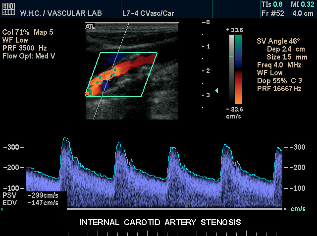
Evaluation of Pulse |
| This color flow doppler US and pulsed doppler interrogation of the neck shows an area of thickening and heaping up off the far wall of the of the internal carotid artery characteristic of atherosclerotic plaque. There is a moderate stenosis of the vessel.
Courtesy Philips Medical Systems. 33301 |
Long Segment Celiac Axis Stenosis with Collaterals from SMA |
|
Angiogram of the SMA shows retrograde filling of the celiac axis which has a significant long segment stenosis. This stenosis has an unusual appearance in that it is relatively long but the most likely cause is atherosclerosis. Courtesy Laura Feldman MD 35076c |
Celiac Axis Stenosis and Subtotal Occlusion of the SMA |
|
The lateral projection of the digital angiogram shows mild to moderate infrarenal atherosclerosis characterised by luminal irregularity. There is also mild lumenal narrowing of the affected infrarenal segment. The suprarenal aorta is smooth and within normal limits. The celiac axis has a high grade sternosis and the SMA shows a subtotal occlusion. Courtesy Laura Feldman MD. 35060 See also 35059 |
|
Narrowing of vessel Caused by Traumatic Spasm |
| This male patient presented with transient loss of pulses and cyanosis of the right foot following traumatic injury to his knee. This angiogram of the popliteal artery, (a, and magnified in b) show spasm and narrowing (red arrow) of the tibioperoneal trunk as well as beading of the artery (white arrows). No obvious intimal injury is identified.
Copyright 2012 Courtesy Ashley Davidoff MD 00008c01.8L |
