The Common Vein Copyright 2012
Definition
Arteritis (Etymology: Latin, arteria, airpipe, itis, inflammation) – is a systemic inflammatory disease usually of large or medium sized arteries. The cause of arteritis is mostly unknown but is believed to often be of an immunological origin. Arteritis has been associated with severe infections and the use of high doses of antibiotics.
Inflammatory change in arteries cause swelling of the walls of the vessels and results in reduced flow to the affected organ . Thus if for example the arteries to the brain are afftected, then the patient may have headaches, visual difficulties, or a stroke.. Systemic signs of inflammation may include malaise, fever, loss of appetite
Imaging studies that may be useful include ultrasound CT scan, MRI and angiography.
The diagnosis of arteritis is confirmed by biopsy.
Corticosteroids is the most common form of treatment.
Polyarteritis Nodosa – Histopathology Occlusive Vascular Disease: Causes of Vascular Occlusion – Polyarteritis Nodosa |
|
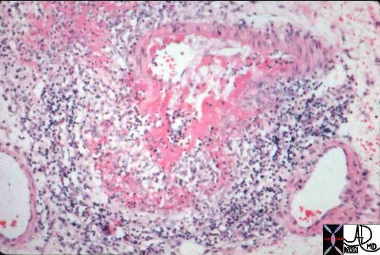
This is a medium power photomicrograph from a bowel in a patient with Polyarteritis Nodosa. Bowel is commonly affected in this disorder. The medium sized artery seen here has undergone a deeply eosinophilic (fibrinoid) change. Notice that there is a surrounding inflammatory infiltrate of WBCs and formation of aneurysmal dilatation. This can result in massive hemorrhage if the aneurysm ruptures. Courtesy Barbara Banner MD 12889 |
Multicentric Vasculitis |
|
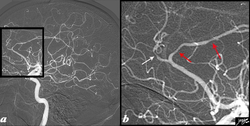
The angiogram is from a 62 year old female patient is with acute neurological deficit The intracerebral angiogram shows focal regions of spasm with one area of spasm in the callosomarginal branch (white arrow) and two foci within the pericallosal artery (red arrows) . Image Courtesy Ashley Davidoff MD Copyright 2012 89028c04.8s |
Multicentric Vasculitis |
|
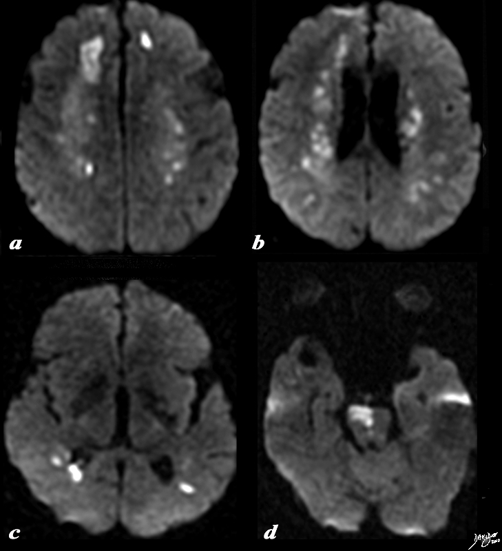
The diffusion weighted MRI is from the 62 year old female patient noted bove who presented with acute neurological deficits. The DWI sequence reveals multicentric of restricted diffusion indicating acute infarction. In image a, the dominant infarction is in the right frontal lobe, but there is an area of infarction in the left frontal lobe as well. Image b shows restricted diffusion in the periventricular white matter, while (c) shows disease in the posterior parietal region. In image (d) an infarction of the right side of the pons is apparent. Image Courtesy Ashley Davidoff MD Copyright 2012 89038c01.8s |
Takayasu’s Arteritis |
|
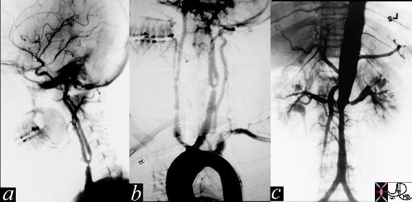
The series of images are from the angiogram of a 14 year old female who presented with seizures and an elevated blood pressure. Images a and b show multiple stenoses within the carotids best seen at the level of the bifurcation into external and internal arteries. In addition in b, the aortic arch shows non critical narrowing just after the origin of the left common carotid vessel. Note that the right subclavian artery is not seen and presumably is accluded at its origin. The abdominal angiogram shows a significant narrowing of the left renal artery with post stenotic dilitation, and stenotic disease in the infrarenal abdominal aorta. The multicentric nature of the disease in a young female is athognomonic of Takayasu’s arteritis. Courtesy of Laura Feldman MD. 35155c |

Multicentric and Diffuse Irregular Narrowing of the Istmus and Abdominal Aorta Takayasu’s Arteritis |
|
An MRI of the thoracoabdominal aorta shows narrowing of the isthmus, distal thoracic segmenet, and the abdominal aorta with both segmental and diffuse changes. The patient has a known diagnosis of Takayasu’s arteritis and aortitis – an inflammatory condition affecting the aorta and large arteries. Copyright 2012 Courtesy Ashley Davidoff MD 16910 |

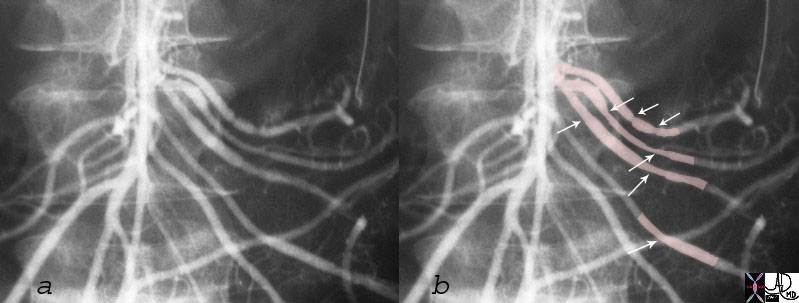
Spastic Change in the Hepatic Artery 2 Months Following Intraarterial Yttrium Therapy Question of Radiation Arteritis |
|
The angiogram of the proper hepatic artery performed in planning for Yttrium therapy (a and magnified in b) in a patient with metastatic carcinoid disease, shows a normal caliber smooth walled right hepatic artery. Hypervascular metastases are suggested in the image (a). The angiogram of the proper hepatic artery performed 2 months later following Yttrium therapy (c and magnified in d) shows beading of the proper (red arrows) and right hepatic artery. These findings raise the possibility of arteritis post Yttrium therapy. Copyright 2012 CourtesyDr Tim Frey113489c01L.8 |
|
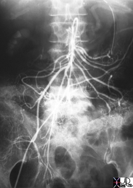
The aortagram is from a 26 year old female with a GI bleed and reveals spastic changes in the jejunal vessels (see deatail images below)
Injection into the superior mesenteric artery (SMA ) revals spastic changes in the jejunal branches, and ileocolic branches. These findings are consistent with a vasculitis.
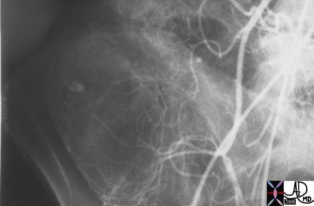
Henoch Schonlein Arteritis presents with an Acute GI bleed |
|
Subsequent subselective injection into the ileocolic artery showed extaravasation characterizing active bleeding into the cecum. Pathology revealed plaque like ulcers in the bowel consistent with chronic ischemia and findings consistent with Henoch -Schonlein arteritis Courtesy Ashley Davidoff MD Copyright 2012 Courtesy Ashley Davidoff MD 28514 28515 28516 28517 surgical specimen showed plaque like ulcers in the bowel consistent with chronic ischemia |