Infarction
Copyright 2009
Definition
Infarction is …
characterized by …..
caused by ..etiology or predisposing factors
resulting in a pathological feature (structural change or functional change) or clinical feauture
Sometimes complicated by ….
Diagnosis is suspected clinically by … and confirmed by ….
Imaging includes the use of
Treatment options depend on …. but includes …..
Etymology if available
Principles
Acute Ischemia

Normal and Global Ischemia after Cardiac Arrest |
| The two images represent a diffusion weighted MRI image which measures Brownian motion of molecules. In acute infarction there is restricted Brownian motion of the affected area and the image can be manipulated to present this as a bright region. In this case the acute infarction or ischemia (b) is relatively bright compared to the white namatter and compared to the gray matterof the normal (a)
49433c02.800 brain cerebral cerebrum white matter gray matter basal ganglia fx increase intensity in gray matter relative decrease in intensity in white matter dx global ischemia question brain death probable irreversible brain death cerebral infarction s/p arrest MRI DWI diffusion weighted imaging normal and abnormal fx diffuse restricted diffusion involving basal ganglia caudate thalami Courtesy Ashley Davidoff MD |
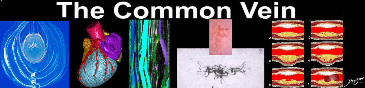
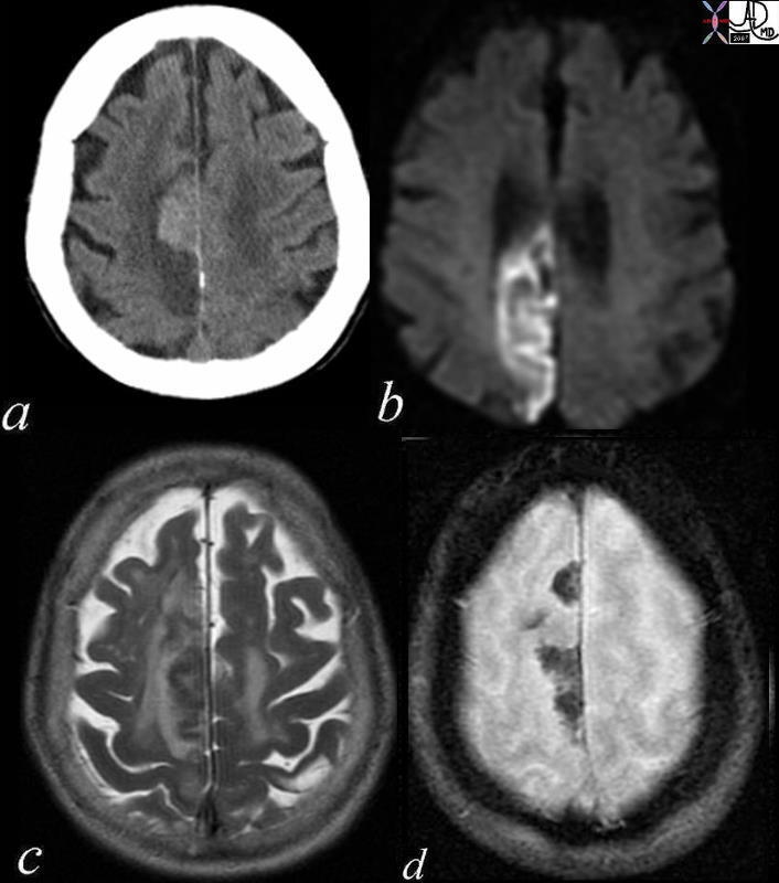
Acute Right Occipital Lobe and Chronic Infarction Left Occiptal Lobe |
| 49678.600 brain occipital lobe fx vague hypodensity right occipital lobe with encephalomalacia and ex vacuo changes in the left occipital and posterior parietal region dx acute infarction right occipital lobe chronic infarction left occipital lobe CTscan Davidoff MD |
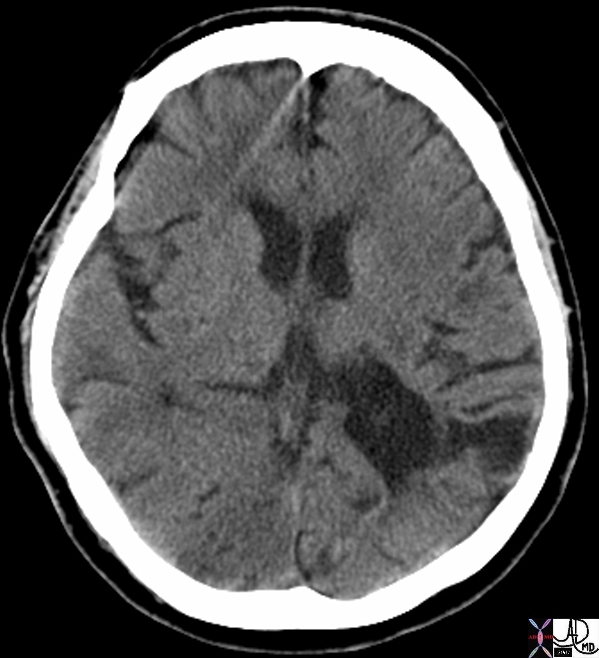
Acute and Chronic Infarction with CT and DWI MRI |
| 49679c01 brain DWI occipital lobe fx vague hypodensity right occipital lobe with encephalomalacia and ex vacuo changes in the left occipital and posterior parietal region dx acute infarction right occipital lobe chronic infarction left occipital lobe CTscan high intesity in right occipital lobe and low intensity in left occipitoparietal region dx acute infarction right occipital lobe chronic infarction left occipital lobe MRI diffusion weighted imaging Courtesy Ashley Davidoff MD |

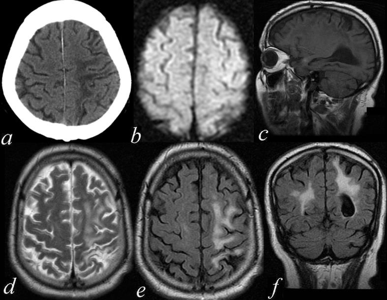
Acute and Chronic Infarction |
| 49685C01 brain DWI occipital lobe fx vague hypodensity right occipital lobe with encephalomalacia and ex vacuo changes in the left occipital and posterior parietal region dx acute infarction right occipital lobe chronic infarction left occipItoparietal lobe a IR white matter disease vague increase in right b T2 gliosis left parietal c DWI bright acute right occipital d CT vague hypodensity right occipital old infarct left dx acute infarction right occipital lobe chronic infarction left parietal lobe MRI diffusion weighted imaging CTscan Courtesy Ashley Davidoff MD |
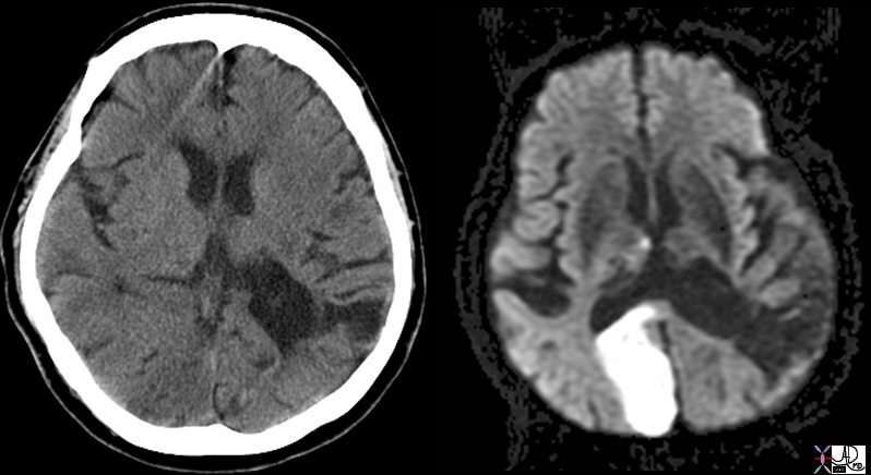
Acute and Chronic by CTscan |
| 71245 brain cerebrum occipital lobe parietal lobe chronic infarction acute infarction hypodensity relative density CTscan Davidoff MD |
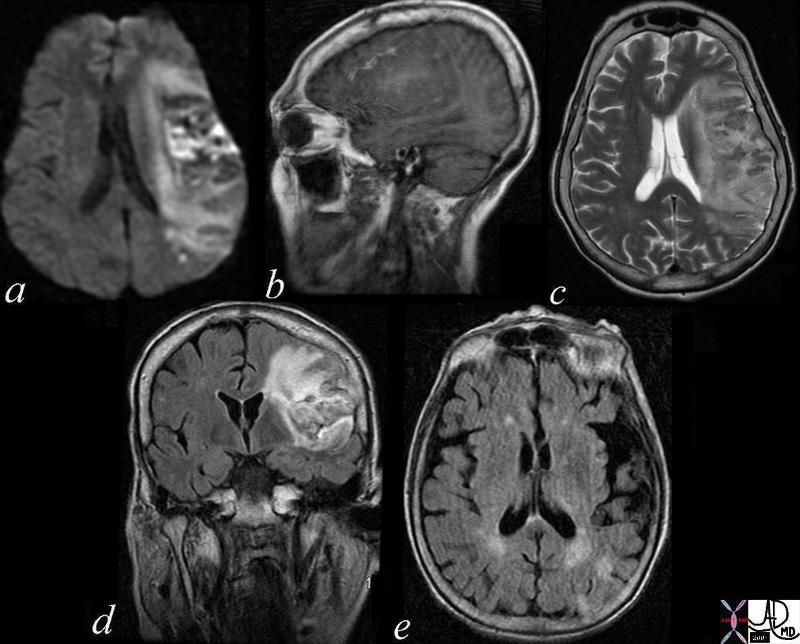
Acute Hemorrhagic Infarction |
| 71239c01 patient with atrial fibrillation brain cerebrum parietal lobe increased density hyperdense parasagittal hemorrhagic infarct paracentral DWII bright T2 bright GRE mixed blood products dx acute hemorrhagic infarction secondary to embolic event the patient disd have other non hemorrhagic infrctions in other parts of the brain CTscan MRI Davidoff MD |
Subacute Infarction
Subacute Hemorrhagic Infarction – 1 month old by history |
| 71000c03 a = DWI b= T1 c= T2 d= FLAIR e=”GRE” 70 Male extensive infarct left MCA territory which has mild mass effect on ventricles petechial hemorrhage T2 and FLAIR hyperintense T2 shine through on diffusion weighted images punctate area in left parietal lobe restricted area of diffusion question recent small infarct MRI about 1 month old from clinical presentation dx subacute hemorrhagic infarct MRI Davidoff MD |
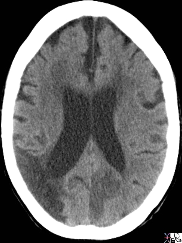
Chronic Infarction
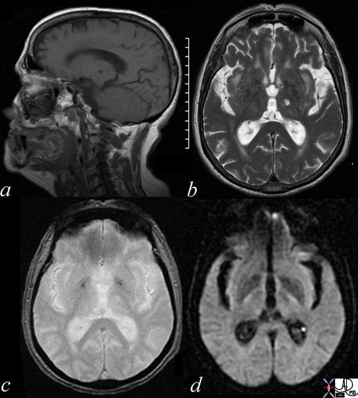
Chronic Thalamic Infarct – a Lacune |
| 70007c01 brain cerebrum cerebral thalamus infarct chronic infarction circulatory MRI sagittal T1- focal hypointense T2 focal hyperintense GRE gradient echo – bright DWI diffusion weighted imaging Davidoff MD |
Chronic Middle Cerebral Artery Infarction |
| 46031c brain cerebrum cerebral fx low density middle cerebral artery territory tempral lobe parietal lobe dx chronic middle cerbral artery infarction with encephalomalacia CTscan |
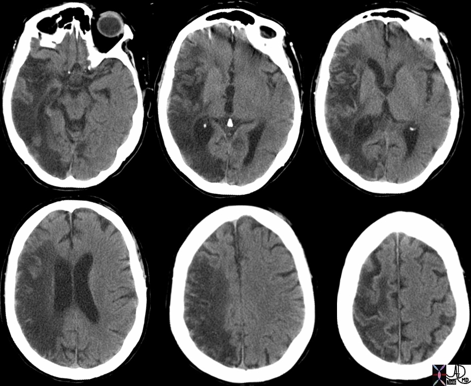
Chronic Ischemic Change in the left MCA territory – Fronto Parietal region |
| 71411c01.800 74 male with mental status changes brain cerebrum cerebral fx gliosis volume loss enlargement lateral ventricle on left encephalomalacia dx superior left frontal lobe parietal lobe in the territory of the left MCA left middle cerebral artery a= CTscan b = DWI – negative c= T1 weighted image d= T2 weighted image e = FLAIR f = FLAIR coronal MRI Davidoff MD |
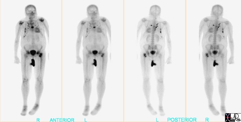
Recent Myocardial Infarction |
| 49688.800 bone heart uptake in ribs right posterior probably related to rib fractures. Finding of interest is the uptake in the heart which should not be seen with a bone scan and it indicates a recent myocrdial infarction NM radioisotope Courtesyt Alan Ashare MD |

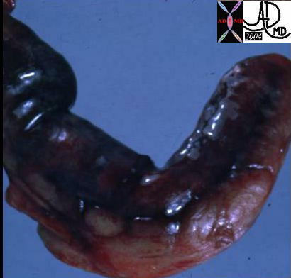
Hemorrhagic Infarct- Subacute |
| Autopsy specimen from a patient with metatsatic lung carcinoma and a hypercoaguable state complicated by DVT, acute portal vein thrombosis and pulmonary embolus (PE), and hemorrhagic pulmonary infarction. The clot noted in the vessels is white, (arrow) and represents subacute thrombus. The vessel remains distended and the distal end of the clot is compacted with the clot. As time passes the clot resorbs, and the vessel returns to nornal size. The wedge shaped defect represents a hemorrhagic infarction
code lungs pulmonary artery thrombosis embolus PE infarction hemorrhage hemorrhagic wedge shaped grosspathology Hamptons hump Courtesy Ashley DAvidoff MD copyright 2009 all rights reserved 32190b01c01.8s |
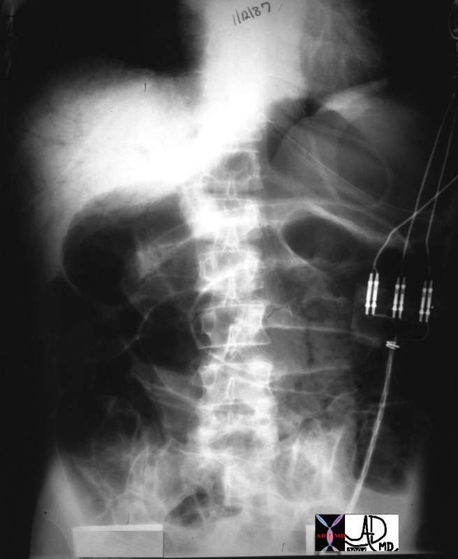
Ischemic Bowel Portal Venous Air |
| 02107 liver hx M52 vein portal fx air small bowel dilated dx ischemiv bowel imaging plain film KUB Courtesy Ashley Davidoff MD DB |
Abscess Complicating Traumatic Infarction of the Liver |
| 24010 liver fx air fx air fluid level loculated air free air dx hepatic abscess following traumatic injury CTscan Courtesy Ashley Davidoff MD |
|
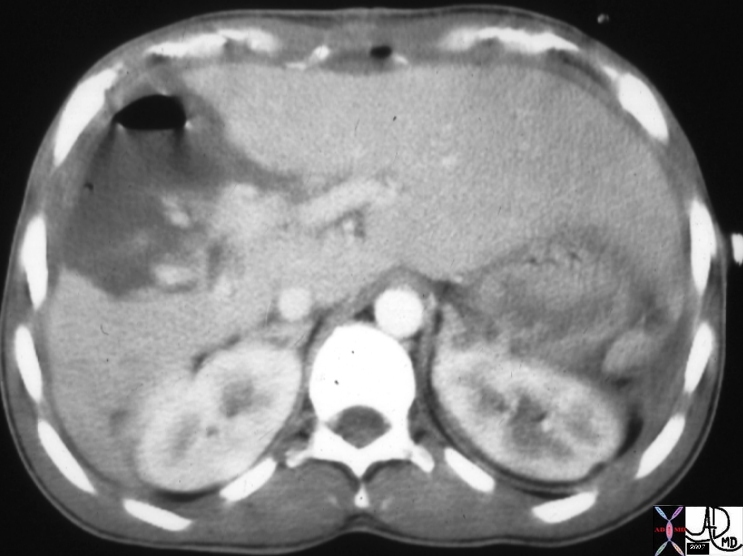
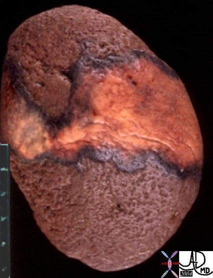
Wedge Shaped Infarct in the Spleen |
| The CTscan with contrast shows a wedge shaped defect in the spleen (overlaid in pink in middle image) characteristic of an acute infarction most commonly caused by emboli. The pathology image from a different patient shows the same wedge shaped process.
46774c01 21321spleen splenic fx wedge shaped defects dx emboli dx systemic embolization dx splenic infarcts CTscan Davidoff MD |

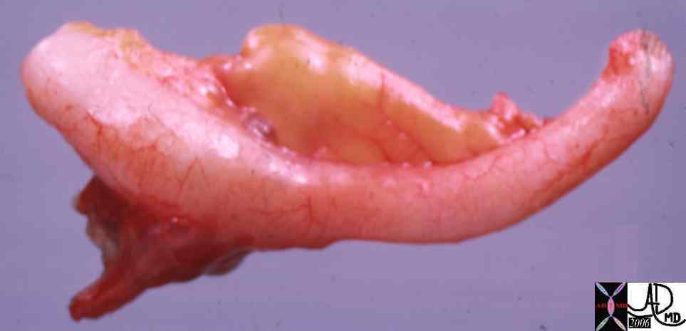 
Normal Lily White Appendix (left) and a Pus Filled Black Necrotic Appendix (right) |
|
02559 colon large bowel appendix grosspathology Courtesy Ted Gulkin MD02435 acute gangrenous appendicitis ( Barbara Banner MD) |
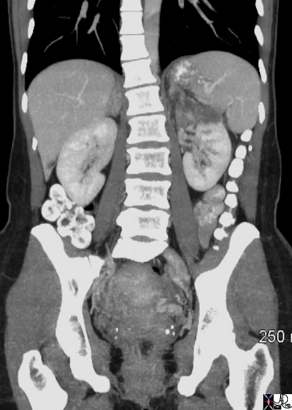 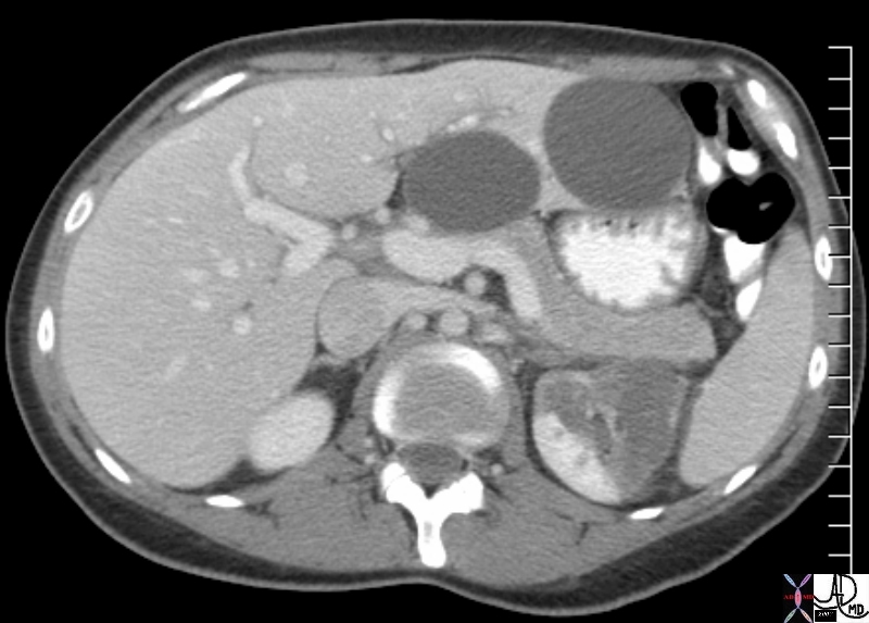
Perfusion Defect with Capsular Enhancement |
| 74263b01 young female presents with pain and white cell count kidney perfusion defect renal luxury perfusion capsular enhancement dx renal infarct vs lobar nephronia infarction pyelopnephritis liver cyst hepatic cyst CTscan Courtesy Ashley Davidoff MD |
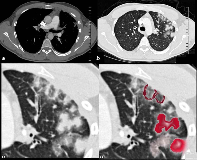
Pulmonary Venous Infarction Secodary to RFA Ablation |
| 75413c06 26 male SOB s/p RFA ablation for atrial fibrillation lung pulmonary let upper lobe pulmonary vein thrombosed secondary lobule interlobular septa are thickened secondary lobule destroyed dx pulmonary vein thrombosis secondary to radiofrequency ablation therapy iatrogenic CTscan Courtesy Ashley Davidoff MD Scott Tsai MD ground glass changes pulmonary infarction venous infarction |