Arteriovenous Malformations
The Common Vein Copyright 2010
Brain
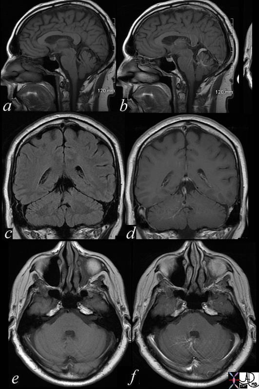
AVM in the Posterior Fossa |
|
brain cerebellum vermis vein enlarged venous angioma old bleed superior left vermis – cerebellar venous malformation vascular malformation a = T1 pre gadolinium b = T1 post contrast c = T1 pre gadolinium d = T1 post contrast e = T1 pre gadolinium f = T1 post contrast MRI Copyright 2012 Courtesy Ashley Davidoff MD 71730c02 |
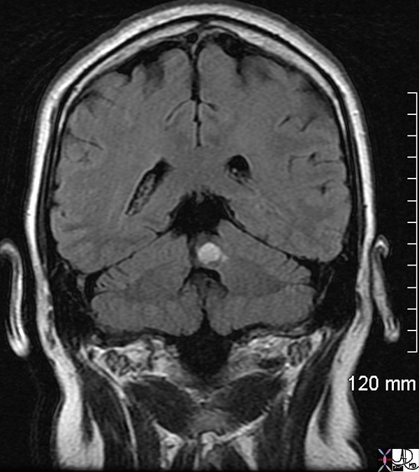
Hemorrhage in an AVM |
|
male with acute ataxia roof of fourth 4th ventricle acute bleed left vermis venous angioma brain cerebellum vermis vein cerebellar venous malformation vascular malformation T1 pre gadolinium T1 bright lesion dx acute hemorrhage MRI Copyright 2012 Courtesy Ashley Davidoff MD 71730 |
Definition
An arteriovenous malformation is a term used to describe a cluster of vessels characterized by the existence of arteries in them connecting directly to veins without there being a capillary bed. They account for approximately 10% of all cerebrovascular malformations.
They are classified according to their blood supply: if it is derived from the internal carotid artery of the vertebrobasilar systems, they are called pial or parenchymal AVM’s. Dural AVM’s are those supplied by the external carotids. They can also be mixed.
Pial arteriovenous malformations usually pose no problem until the main issue associated with them arises: spontaneous hemorrhage, with an eventual associated seizure. The hemorrhage is intraventricular or parenchymal, and is as described in its corresponding review. The risk for bleeding is 2-3% per year.
As for patients with dural AVM’s, they present with complains derived from a fistula in the territory of the external carotid system – pulsatile tinnitus, bruits, headaches, facial spasms.
Diagnosis is made thorough imaging. They are best seen using MRI; however CT can show small calcifications that are associated with these formations. With contrast, it will show typical vascular channels, mostly in the supratentorial area where they are more frequent. They appear as a nidus of enlarged vessels with entangled loops, having no mass effect. MR demonstrates complex flow patterns, with dark areas in T2WI. 3D time of flight sequences can be used to image the fast-flow components. 10% of AVMs develop an associated aneurysm, usually on the feeding artery. Conventional angiography easily diagnoses large AVM’s, and is used for the characterization and delineation of these formations.
Treatment is done with interventional radiological procedures, involving basically embolization or radiosurgery, though direct surgical interventions are also used.
|
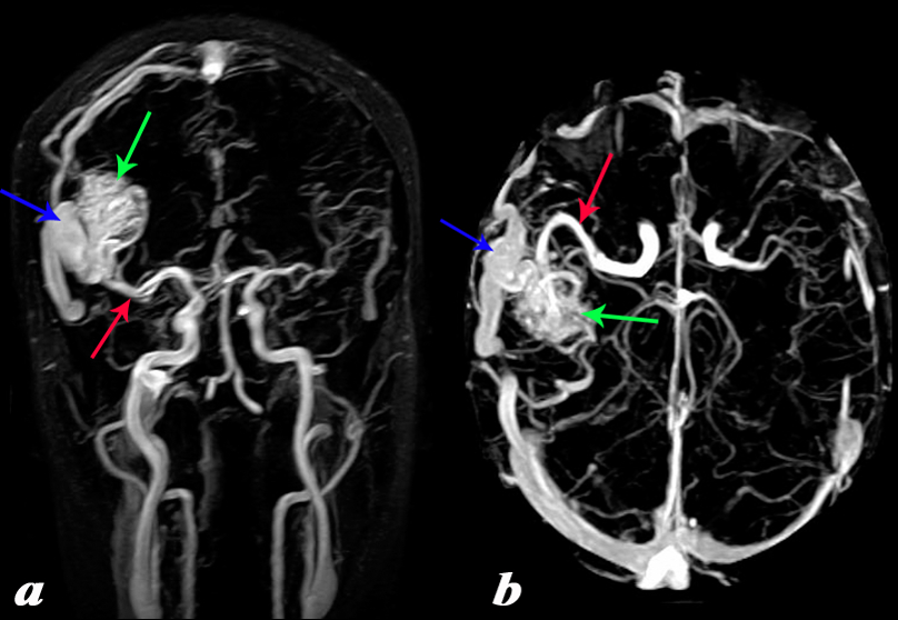
Temperoparietal AVM Large Feeding Artery and Large Draining Vein |
|
The MRA with 2d TOF in the coronal projection (a) and in the axial projection(b). It shows an arteriovenous malformation (AVM) in the right temperoparietal region fed by an enlarged middle cerebral artery (red arrow) and draining into a large draining vein (blue arrow) resulting in an enlarged transverse sinus. Note the difference in size between the right and left transverse sinuses. Courtesy Philips Medical Systems 92465c01L.8 |
|
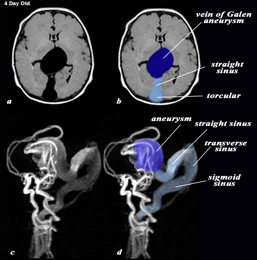
Vein of Galen Aneurysm – 4day Old with AVM |
|
The MRI with a T1 weighted axial sequence (a,b) and with 3d TOF (c,d) shows a large aneurysm of the vein of Galen (dark blue (b,d) an enlarged straight sinus (light blue b) and enlarged draining transverse sinus and sigmoid sinus (c,d) The feeding vessels of the arteriovenous malformation (AVM) which is the cause of the aneurysm, seem to arise predominantly from the vertebro-basilar system and likely from the posterior cerebral artery, but the middle cerebral artery also appears to contribute to the AVM. Courtesy Philips Medical Systems 92432cL.9 |
|
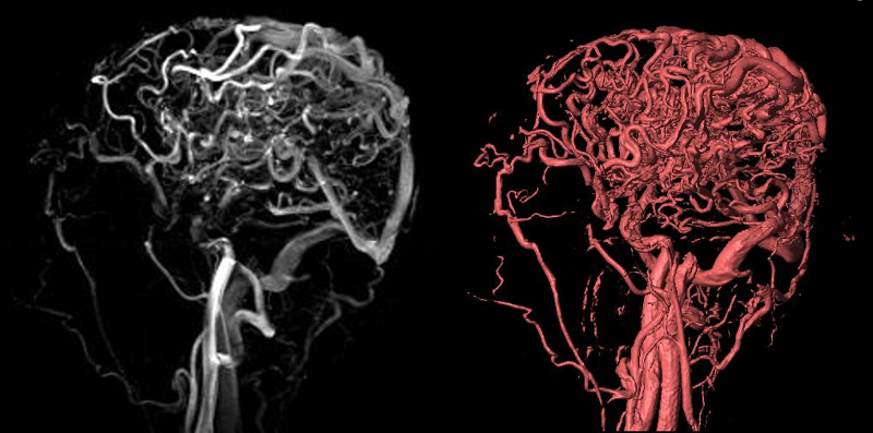
Extensive Cerebral Varicocities – AVM Caput Medusa |
|
Exetnsive varicocities are seen in the parieto-occipital regionin the 2D TOF and volume rendered image. The draining transverse sinus and sigmoid sinus are also enlarged. Courtesy Philips Medical Systems 92455c.8 |