The Common Vein Copyright 2010
Definiton

Acute Hemorrhagic Infarction |
|
The brain has a characteristic appearance characterised by its creamy color and characteristic shape and fissured surface. This pathological specimen shows an acute hemorrhagic infarct in the left frontoparietal region with necrosis of the brain tissue and mass effect on the lateral ventricle and midline shift. The specimen also serves to reveal the normal right side with its creamy color and darker brownish gray matter that is accentuated by increasing the contrast on the right image (b). Other structures that are seen include the corpus callosum that lies above the ventricles (cc), the caudate nucleus (c) that lies inferior to the lateral ventricle, the line of white matter between the caudate and putamen called the internal capsule, the 3rd ventricle (3) medial to these structures , and the thalamus (th) inferior to them. The interhemispheric fissure (if) is seen superiorly and the Sylvian fissure (Sf) is seen laterally. The horizontal folds and darker color of the cerebellum are characteristics of that structure. Courtesy Ashley Davidoff MD Copyright 2010 10407c01 |
Imaging
An acute infarct of the brain is characterised by a focal hypodense area, in cortical, subcortical, or deep gray or white matter, following vascular territory, or watershed distribution. Early subtle findings include obscuration of gray/white matter interface, effacement of sulci, or “insular ribbon.”
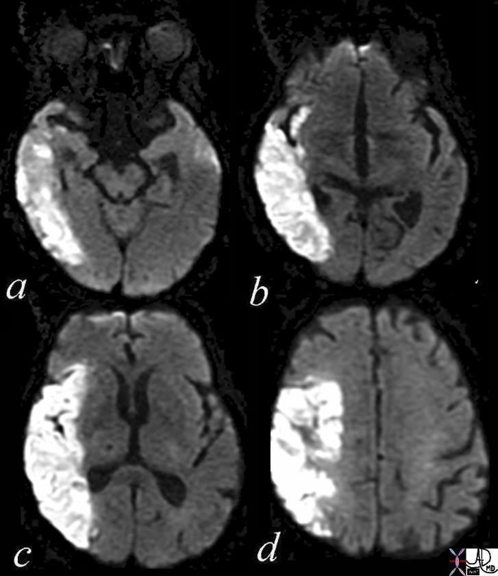
Acute Infarction Parietal and Temporal Lobe DWI |
|
The diffusion weighted MRI in axial projection shows a high intensity region in the right parietal and right temporal region revealing an acute infarction poattern in these regions reflecting right middle cerebral artery territory. Note that the basal ganglia have been spared, but the insula and operculum are involved. Courtesy Ashley Davidoff MD Copyright 2010 71275c01c03 |
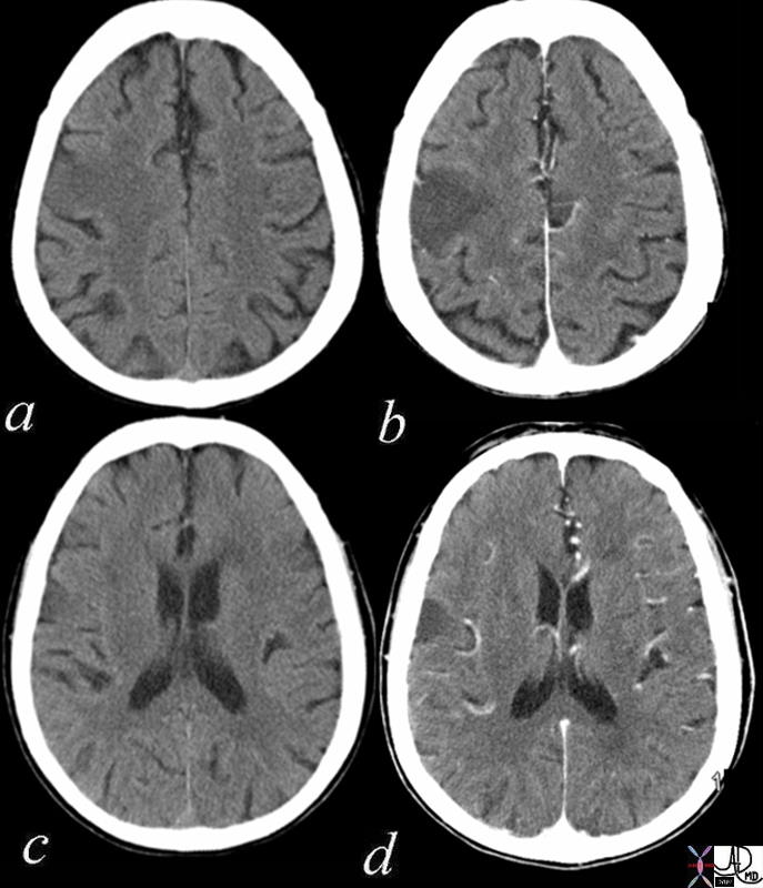
Acute Infarct Right Frontoparietal Region |
|
The axial CT scan through the ventricles is from a 73year old female with acute left sided weakness and slurred speech. Images a nd c are without contrast and images b and d are following intravenous contrast. In the right frontoparietal region within the location of the pre and post central gyrus there is a focal hypodense region accentuated in the contrast study, with loss of the gray white differentiation, consistent with an acute infarction in the parietal lobe. Courtesy Ashley Davidoff MD Copyright 2010 71253c01 |
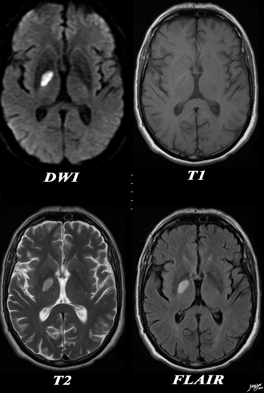
Acute Infarction – Right Globus Pallidus |
|
The basal ganglia with an abnormality in the right globus pallidus are shown in axial projection in the MRI of a 64 year old male who presents with acute neurological deficit. In the first image a high intensity region in the right globus pallidus is shown in axial projection on DWI consistent with an acute infarction. The second image is a T1 weighted image without contrast and shows a rim of high intensity suggesting rim hemorrhage.. The third T2 weighted image shows the lesion to be bright but not as bright as CSF a finding consistent with the acute infarction. The fourth image is a FLAIR image and also shows increased intensity of the lesion. The constellation of findings is consistent with an acute infarction of the right globus pallidus with a rim of acute hemorrhage. Courtesy Ashley Davidoff MD Copyright 2010 All rights reserved 89087c01.8s |
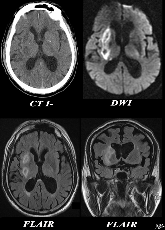
Acute Thalamostriate Infarction |
|
The CT and MRI images are from an 82 year old make with acute neurological deficit. An acute infarction is shown that involves territory within and beyond the basal ganglia. In the CTscan two low density regions are seen medial to the Sylvian fissure and medial to the insular cortex, likely involving the putamen and the part of the right caudate nucleus as well as some white matter in right middle cerebra;l territory. In the second image a high intensity region in the putamen and part of the caudate nucleus is shown together with an area that does not involve the basal ganglia more posteriorly, likely part of the white matter of the right parietal lobe shown in an axial projection on DWI consistent with an acute infarction. The third image is an axial FLAIR sequence and shows the area of parenchymal edema with presumed central necrosis, while the image at low right shows the periventricular disease to better advantage. The constellation of findings is consistent with an acute infarction with distribution of the lenticulostriate vessels arising from the right middle cerebral territory Courtesy Ashley Davidoff MD Copyright 2010 All rights reserved 89367c01.8s |
|
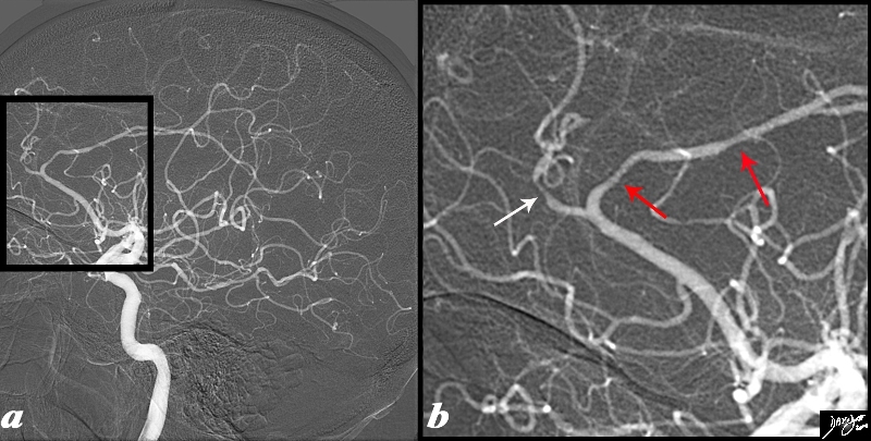
Vasospasm in the Anterior Cerebral Artery – Vasculitis |
|
The angiogram is from a 62 year old female patient is with acute neurological deficit The intracerebral angiogram shows focal regions of spasm with one area of spasm in the callosomarginal branch (white arrow) and two foci within the pericallosal artery (red arrows) . Image Ashley Davidoff MD Copyright 2010 89028c04.8s |
|
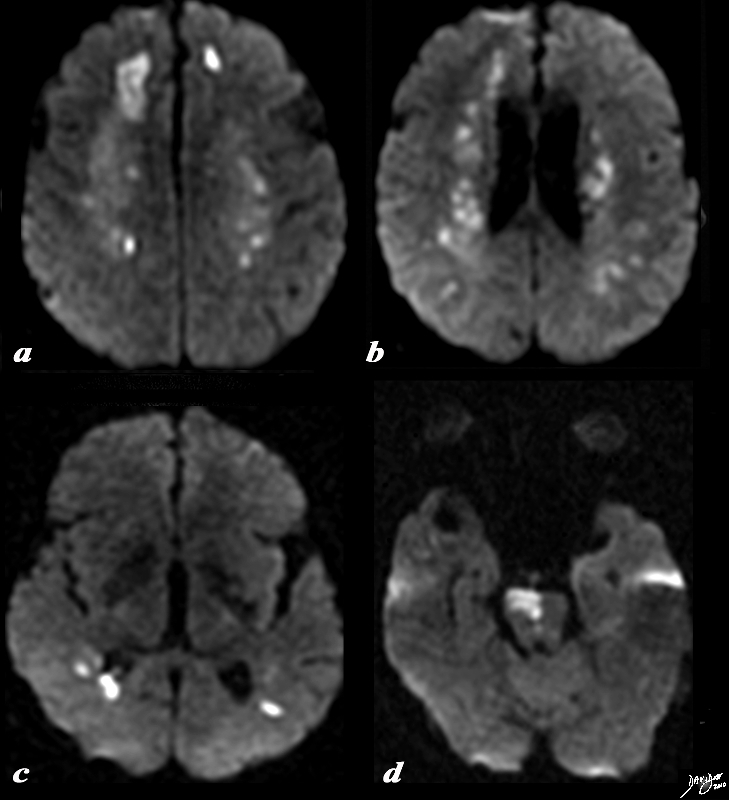
DWI – Multicentric Infarction – Acute Vasculitis |
|
The diffusion weighted MRI is from a 62 year old female patient is with acute neurological deficit The DWI sequence reveals multicentric of restricted diffusion indicating acute infarction. In image a, the dominant infarction is in the right frontal lobe, but there is an area of infarction in the left frontal lobe as well. Image b shows restricted diffusion in the periventricular white matter, while (c) shows disease in the posterior parietal region. In image (d) an infarction of the right side of the pons is apparent. Courtesy Ashley Davidoff MD Copyright 2010 89038c01.8s |
|
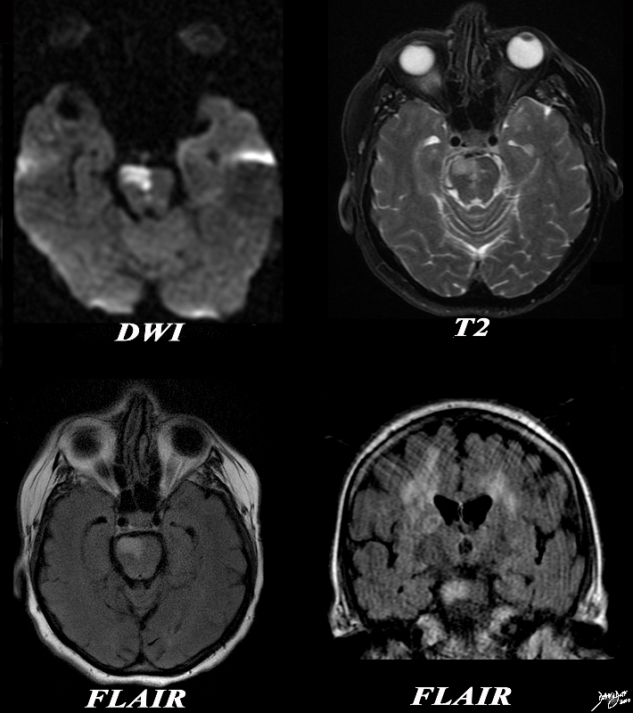
Acute Infarction in the Pons – Vasculitis |
|
The diffusion weighted MRI is from a 62 year old female patient is with acute neurological deficit The DWI sequence in image (a) reveals a focus of restricted diffusion in the pons. This finding is confirmed on the T 2weighted image (b) as well as on FLAIR on the axial FLAIR (c) and coronal FLAIR(d). On the last image – the coronal image shows FLAIR hyperintensity dominant in the right white matter particularly in the corona radiate. These findings are consistent with an acute infarction in the pons and the multicentricty is compatible with the diagnosis of vasculitis. Image Ashley Davidoff MD Copyright 2010 89038c.8s |
|
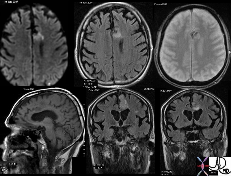
Acute Hemorrhagic Infarction |
|
The MRI in various projections confirms the presence of an acute hemorrhagic infarct in the left paracentral frontal territory. The diffusion images (top left), show a hyperintense focus in the left frontal lobe, which is hyperintense on the axial FLAIR image (middle top), hypointense and granular on the GRE (top right), hypointense on the sagittal T1 (bottom left) and seen as hyperintense on the last two STIR coronal images. These findings are consistent with an acute hemorrhagic focus which and was thought to be embolic in origin due to other noted foci. Courtesy Ashley Davidoff MD copyright 2010 46098c01.800 |