Cerebral Aneurysms
The Common Vein Copyright 2010
Definition
Cerebral aneurysms denote a condition in which an artery that irrigates the brain is abnormally dilated. These can occur in any of these arteries, and are commonly divided in those belonging to the anterior circulation (that is, from the internal carotid or its branches) or those belonging to the posterior circulation (from the vertebrobasilar system and corresponding branches).
They are caused in part by degeneration of the wall of the arteries secondary to hemodynamic stress. As such, hypertension and smoking are risk factors that predispose to the development of these aneurysms, which directly or indirectly increase this stress. Connective tissue diseases, the presence of arteriovenous malformations and polycystic renal disease are also connoted with this condition.
They vary in size and shape. They are classified according to their etiology and shape; initially they are divided into saccular and nonsaccular types. The former (also called berry) typically arise at a bifurcation of a vessel; The nonsaccular types can be atherosclerotic, fusiform, mycotic and traumatic.
They can be asymptomatic or have serious consequences, particularly if they rupture (since the wall becomes weaker), giving rise to subarachnoid hemorrhages. Size are important to determine risk of bleeding, that usually becomes significant for sizes over 5 mm. When a rupture occurs, dramatic symptoms develop, with a sudden excruciating headache as described in the SAH review.
Radiologically, MRI is able to provide details about the aneurysms, with the above mentioned characteristics: size, shape and content. These details are significantly improved after administration of IV contrast as it might be expected. MRA is very sensitive in population with increased risk. Treatment is surgically, and prior to the procedure, angiography is performed to add significant data such as collateral circulation, associated vascular diseases and cerebral vasospasm. Interventional radiology has also developed treatment for this condition and carries tools that allow clipping, introduction of coils and balloon angioplasties.
Anterior Communicating Aneurysms
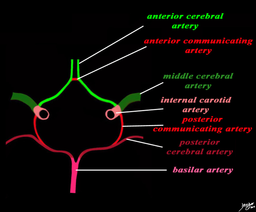 Anterior Communicating Artery Anterior Communicating Artery
A tiny but essential component in the circle of Willis |
|
The diagram shows the main branches of the blood supply to the brain which includes the carotid and vertebro-basilar systems. These are the vessels that particpate in the formationofthe circle of Willis The carotid system supplies the brain from the internal carotid (salmon pink) – a branch of the common carotid which arises from the aorta. Its terminal bifuracation into the middle cerebral (dark green) and anterior green (bright green) are shown. The anterior communicating artery runs between the two anterior cerebrals (bright red) The basilar artery (pink) is formed by the two vertebral arteries and travel as a single artery over the upper medulla and the entire pons. Its terminal branch is the posterior cerebral artery (maroon). Each of the vessels contributes to the circle of Willis through communicating arteries. The vertebro-basilar system provides the posterior communicating arteries bilaterally and the carotid system provides the anterior communicating arteries via the anterior cerebral artery. code brain Courtesy Ashley Davidoff MD Copyright 2010 All rights reserved 97194b13g04L01.91s |
|
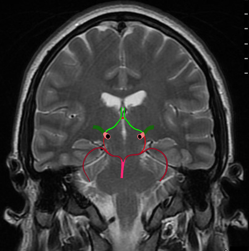
The Circle of Willis Overlaid on a Coronal View of the Brain |
|
The diagram shows the main branches of the blood supply to the brain including the circle of Willis overlaid on coronal MRI image to portray the approximate position of the vessels in the brain. The carotid system supplies the brain from the internal carotid we demonstrate its terminal bifurcation into middle cerebral (dark green) and anterior cerebral (bright green). The anterior communicating artery runs between the two anterior cerebrals (bright red) The basilar artery (pink) is formed by the two vertebral arteries and it travels as a single artery over the upper medulla and the pons. Its terminal branch is the posterior cerebral artery (maroon). Each of the carotid and vertebro-basilar systems contributes to the circle of Willis through communicating arteries. The vertebro-basilar system provides the posterior communicating arteries bilaterally from the posterior cerebral and the carotid system provides the anterior communicating arteries via the anterior cerebral arteries. Courtesy Ashley Davidoff MD Copyright 2010 All rights reserved 89721c06b.8sg04.8s |
Anterior Communicating Artery Aneurysm
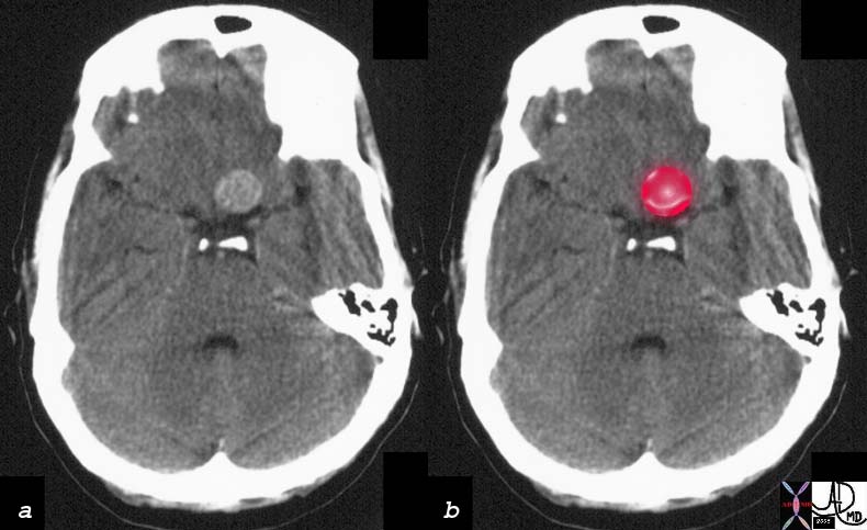 Aneurysm of the Anterior Communicating Artery Aneurysm of the Anterior Communicating Artery |
|
The CT with contrast in axial projection shows an enhancing aneurysm in the region of the circle of Willis that was shown to arise from the anterior communicating artery. See angiogram below Courtesy Ashley Davidoff MD 16004c01 |
|
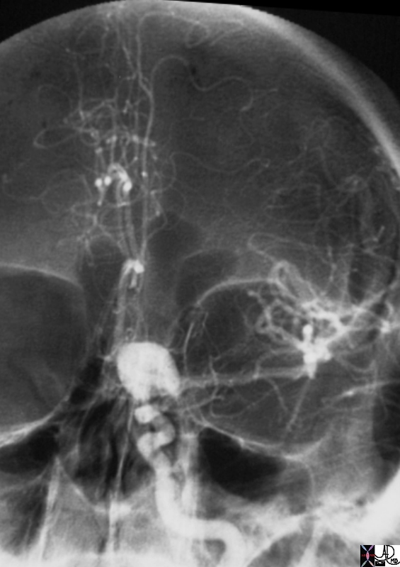
Anterior Communicating Artery Aneurysm |
|
The angiogram in the A-P projection shows the aneurysm in the region of the circle of Willis. The aneurysm arose from the anterior communicating artery. The middle cerebral artery appears normal. Both anterior cerebral arteries fill suggesting that the anterior communicating artery is patent. 16006.800 Courtesy Ashley Davidoff MD |
|
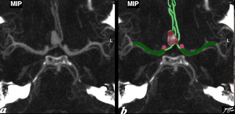
Anterior Communicating Artery Aneurysm – CTA |
|
The CTA through the circle of Willis shows an aneurysm in close association with the anterior cerebral arteries (bright green). It appears to lie between the anterior cerebral arteries and likely represents an aneurysm of the anterior communicating artery. The distal internal carotid is seen in salmon pink while the middle cerebral artery is overlaid in dark green. Image courtesy Philips medical Systems Image rendering Ashley Davidoff MD Copyright 2010 92577c01b8 |
|
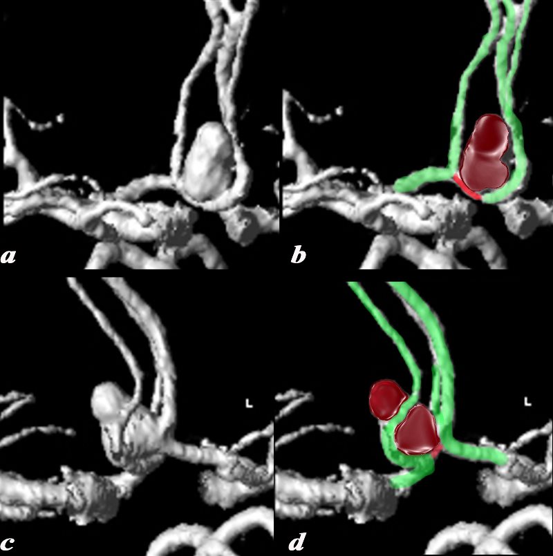
3D Volume Rendering of the Aneurysm Enabling Identification of the Anterior Communicating Artery |
|
The CTA through the circle of Willis with 3D volume rendering shows an aneurysm in close association with the anterior cerebral arteries (bright green). These views allow the visualization of the anterior communicating artery (red) confirming that the origin of the aneurysm is in fact from the anterior communicating artery. The 3D views allow more definitive evaluation of the origin of the aneurysm Image courtesy Philips medical Systems Image rendering Ashley Davidoff MD Copyright 2010 92577c04.8s |
|
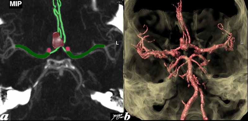
Aneurysm in Contextual Perspective |
|
The CTA through the circle of Willis shows an aneurysm in close association with the anterior cerebral arteries (bright green). It appears to lie between the anterior cerebral arteries and likely represents an aneurysm of the anterior communicating artery. The distal internal carotid is seen in salmon pink while the middle cerebral artery is overlaid in dark green. Image b shows 3D volume rendered anterior and posterior circulation in the context of the base of the skull giving a contextual perspective of the aneurysm. Image courtesy Philips medical Systems Image rendering Ashley Davidoff MD Copyright 2010 92577c.81 |
MCA Aneurysm
|
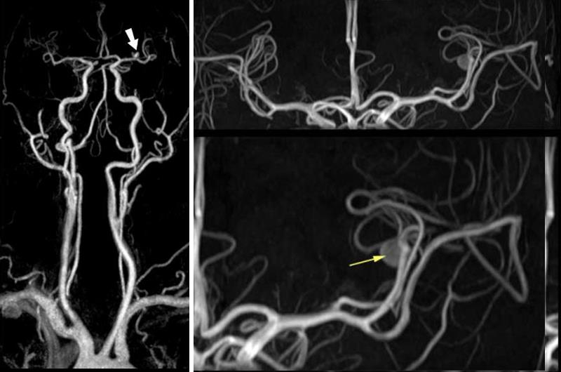
MRA – Left MCA Aneurysm |
|
The MRA performed on a 3T magnet shows an aneurysm of the left middle cerebral artery (MCA) demonstrated in the global frontal view (left image) by a large white arrow, and in the magnified view of the MCA (yellow arrow) Image Courtesy of Philips Medical Systems 92812.8 |
|
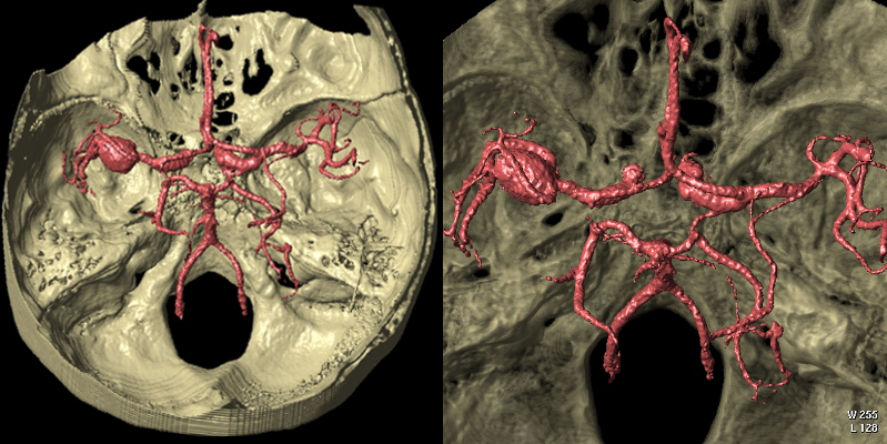
Right MCA Aneurysm and Ectasia of MCA’s Bilaterally CT – Volume Rendering |
|
The 3 D volume rendered image of the cerebral circulation looking into the middle and posterior cranial fossa with the circle of Willis in view shows a right sided middle cerebral artery aneurysm. Both middle cerebral arteries appear ectatic as well. Courtesy Philips Medical Systems 92579c |
Aneurysm of the Basilar Artery
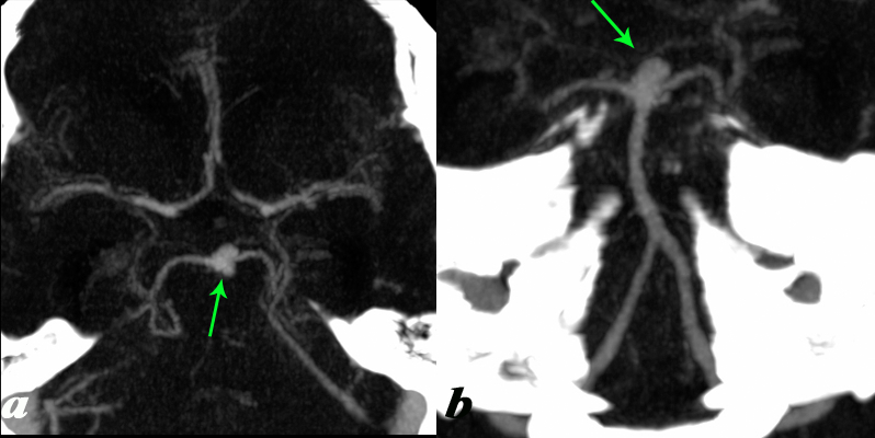 Basilar Tip Aneurysm Basilar Tip Aneurysm |
|
The axial (a) and coronal (b) image of the vertebro- basilar circulation looking into the middle and posterior cranial fossa with the circle of Willis in view shows a basilar tip aneurysm. Courtesy Philips Medical Systems 92584c |
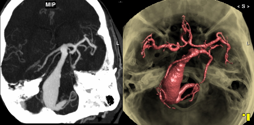 Fusiform Aneurysm of the Basilar Artery Fusiform Aneurysm of the Basilar Artery |
|
The MIP (left) and 3D volume rendered (right) image of the vertebro- basilar circulation looking into the middle and posterior cranial fossa shows a diffuse fusiform aneurysm of the basilar artery. Courtesy Philips Medical Systems 92415c.8 |
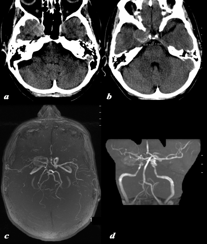 Aneurysm in the Region of the Right ICA Bifurcation Aneurysm in the Region of the Right ICA Bifurcation |
|
The CT scan of the head is from a 62 year old female who presented with right eye paralysis. The CT (a,b) shows a 21mmX 16mm mass on the right medial temporal surface The MRA in stacked axial (c) and coronal (d) projection shows an incompletely filled aneurysm likely arising from the distal supraclinoid portion of the internal carotid or close to the bifurcation. These studies are not sufficiently precise to enable the distinction. Image Courtesy Ashley Davidoff MD Copyright 2010 All rights reserved 88985c.8L |
 Broad Based Internal Carotid Artery Aneurysm Broad Based Internal Carotid Artery Aneurysm |
|
The selective right common carotid angiogram shows a small and broad based internal carotid artery aneurysm (purple) arising form the supraclinoid portion (salmon pink). The ophthalmic artery (red) that arises from the border between the supraclinoid portion and the cavernous portion of the internal carotid artery is noted Other components noted include the cavernous portion (orange), the division into the anterior cerebral arteries, (bright green) and middle cerebral artery (dark green). Image Courtesy Ram Chavalli MD Copyright 2010 All rights reserved 49409Sc01.8 |