The Common Vein Copyright 2010
Definition
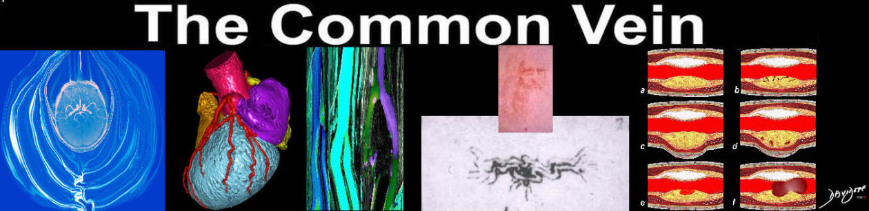
Hypertonicity of a Chronic Infarction |
|
60 year old malewith The classical appearance of hypertonia of upper limb and hand muscles is shown in this CT volume rendered image. The hand and elbow are flexed as a result of involvement on the contralateral motor cortex. Courtesy Ashley Davidoff MD 48641c01.800 |
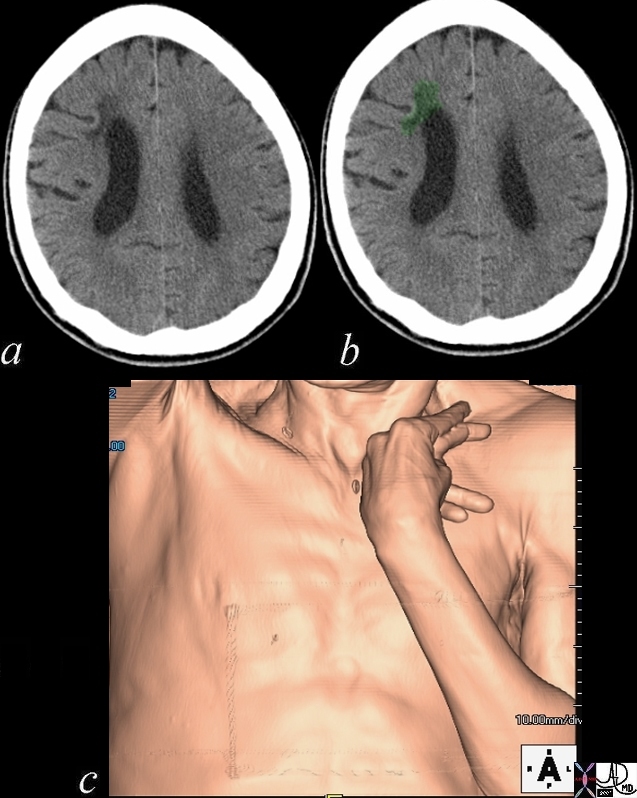
Chronic Bilateral Infarcts |
| The axial CT through the inferior aspect of the frontal lobes show blateral low density regions in the frontal lobes with volume loss indicative of chronic infarcts bilaterally with encephalomalacia
Courtesy Ashley Davidoff MD Copyright 2010 71404 |
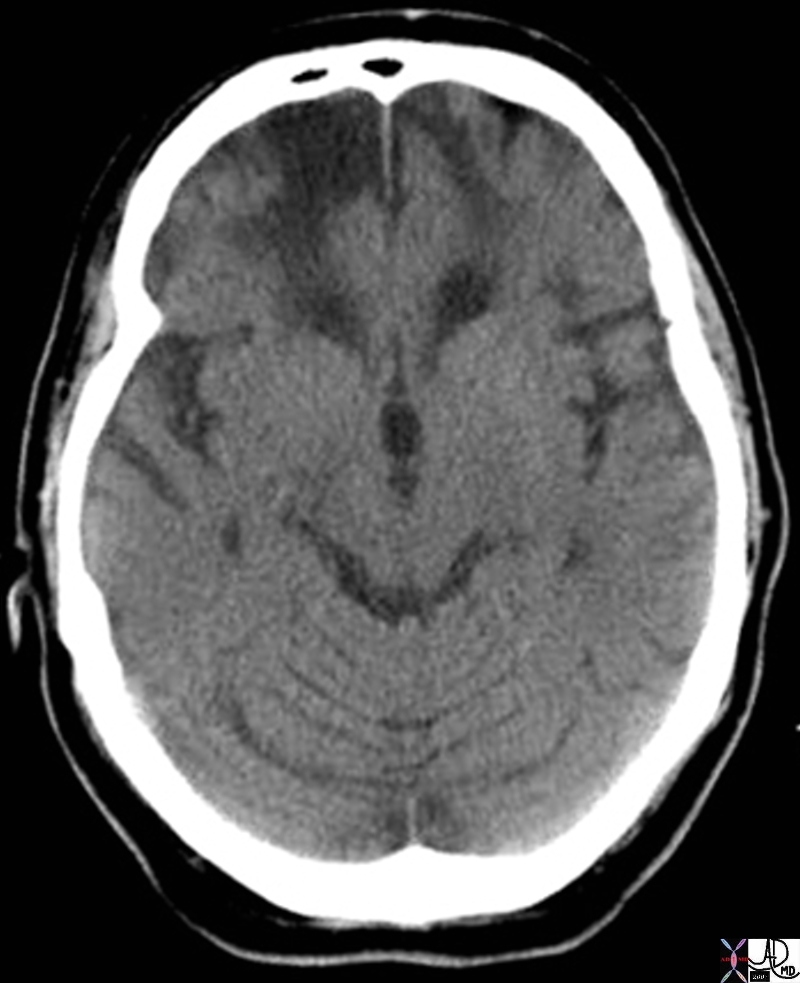
Chroni Parietal Infarction |
|
The CT scan shows the effects of an extensive right middle cerebral infarct and involves the right parietal lobe, temporal lobe, and occipital lobe. The right lateral ventricle is enlarged because of the loss of brain tissue as a result of the infarction. This process is called encephalomalacia (brain softening) and the changes are called ex vacuo changes because the loss of tissue results in gain of space resulting in the shift of the ventricle into the space. Courtesy Ashley Davidoff MD copyright 2010 46031c |
|
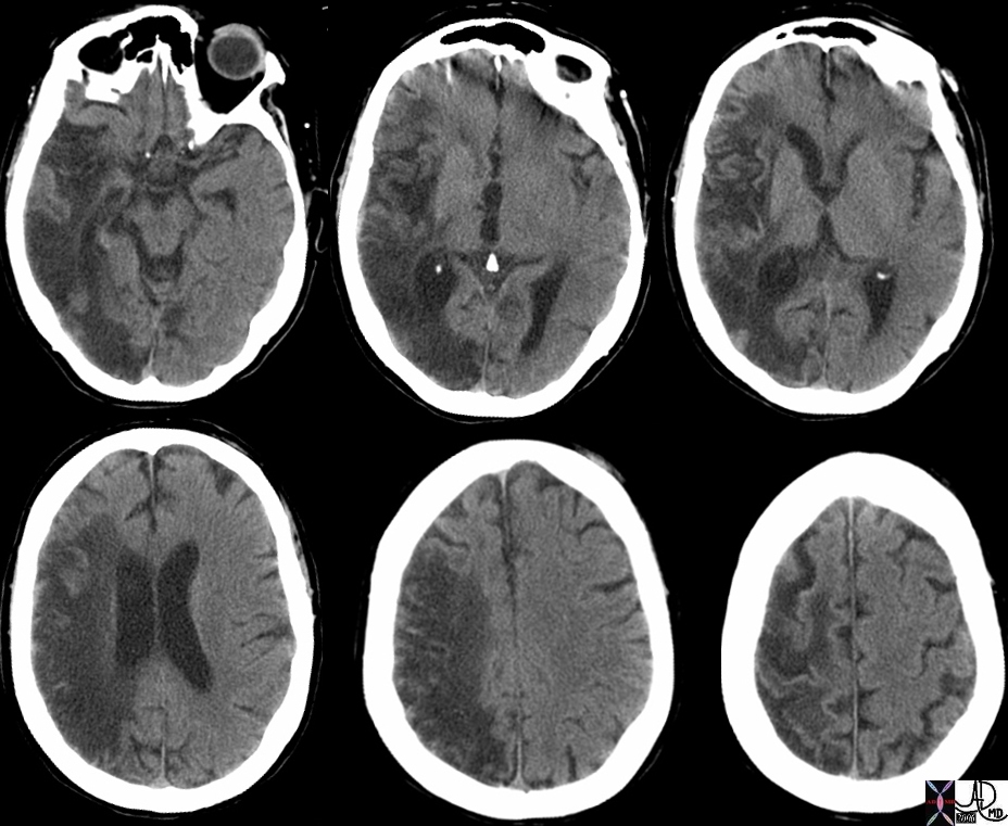
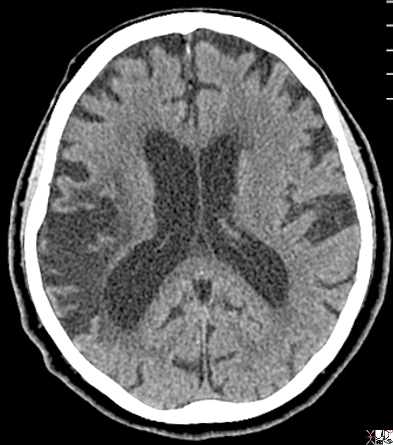
Encephalomalacia, Ex Vacuo Change and Asymmetric Enlargement of the Posterior Horn |
|
The CT scan of this 94 year old patient reveals asymmetry in the occipital horns caused by volume loss of the posterior right parietal lobe as a result of a chronic right middle cerebral infarction. The posterior horn of the ventricle has expanded to fill the space left by the encephalomalacia as a result of the infarction. This is called ex vacuo change and results in asymmetric enlargement of the ipsilateral ventricle. Courtesy Ashley Davidoff MD Copyright 2010 89331b.8s |
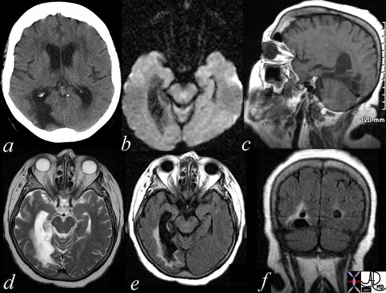
Chronic Infarction – Posterior Cerebral Circulation |
|
The patient is a 74 year old female with a history of ataxia, incontinence, and memory loss. The MRI shows features of a chronic infarction in the right posterior cerebral artery territory affecting the posteromedial aspect of right temporal lobe and medial and inferior aspect of the right occipital lobe. There is evidence of gliosis with volume loss, atrophy, and encephalomalacia resulting in ex vacuo changes with dilatation of temporal and occipital horns of the lateral ventricles. Image a is a CTscan showing encephalomalacia in the right posterior temporal region and ex vacuo change with ventricular dilatation. Image b is a DWI scan showing no acute changes. Image c is a T1 weighted sagittal view showing no acute hemorrhage. Image d is a T2 weighted study showing compound high intensity changes in the region of the occipital horn but the distinction between disease and dilated ventricle cannot be made because of blooming and isointensity of the ventrcles and the diseases cerebral tissue. Image e is a FLAIR image that turns the CSF black and enables the identification of the high intensity abnormal brain tissue that is smaller than expected after reviewing the T2 weighted image. This case reveals the importance of the FLAIR image. Image f is a coronal FLAIR sequence also showing that the actual dimension of diseased tissue is relatively small. The findings are consistent with an old chronic infarction. Courtesy Ashley Davidoff MD Copyright 2010 All rights reserved 71419c01.800 |