VENOUS DRAINAGE
The final common pathway of hepatic venous drainage is via the three hepatic veins which enter into the IVC at the dome of the liver.
Tributaries include the right hepatic vein, middle hepatic vein, and left hepatic vein.
The veins are formed by branches of the segmental and subsegmental veins of the liver, and are originally formed at the center of the liver lobule where the hepatic venous radicle is called the central vein.
It is called the central vein because the sinusoids formed by the terminal portal venous and arterial terminal branches, converge to this central point of the liver lobule before they finally drain out of the liver.
The venous drainage of the caudate lobe enters independently into the IVC at a point more caudal than the major hepatic veins.
The hepatic venous anatomy forms the boundaries of the lobes and segments of the liver.
| The hepatic veins originate from the sinusoids and convey blood from the liver to the vena cava. Hepatic veins tend to be open and solitary, allowing them to be distinguished from the branches of the portal vein, which are more or less collapsed and always accompanied by an artery and duct.

Lobule |
|
A branch of the hepatic vein terminating in a liver lobule. (Image courtesy of Ashley Davidoff M.D.)
|

Lobules |
|
There are a multiplicity of lobules, each with a central vein and each delivering the “goods” to venules, which collectively join to form the hepatic veins and then into the IVC. Destination? The heart, from where it will be distributed to the body wide system. (Image courtesy of Ashley Davidoff M.D.)
|

Venogram |
|
The venogram shows a catheter in the middle hepatic vein filling the venules of a subsegment. The destination of the blood is the inferior vena cava, which is the structure filled with contrast above the diaphragm. (red color overlay) (Image courtesy of Ashley Davidoff M.D.)
|

Bunny Ears |
|
This cross sectional ultrasound image of the confluence of the hepatic veins has been likened to the face of a “bunny”. (Image courtesy of Ashley Davidoff M.D.)
|

Hepatic Veins |
|
This coronal view of the abdominal cavity shows the confluence of the 3 major hepatic veins as they form the IVC at the diaphragm. (Image courtesy of Ashley Davidoff M.D.)
The hepatic vein Is a medium sized vein structurally characterised by its position in the cranial portion of the liver and its connection to the ivc functionally characterised by being the final confluens of venous drainage from the lobes arising from the segmental veins entering into the ivc common diseases include congestion clinical presentation RUQ pain due to congestion large liver diagnostic studies include US, CT, MRI treatment is commonly by medical therapy
|

Position and Direction of Blood Flow |
| 46538c01 liver hepatic veins acv waves normal anatomy physiology venous drainage Dopppler flow color flo direction USscan Davidoff MD |
Applied biology
Caput Medusa is a clinical sign characterised by prominent periumbilical veins, reminiscent of the Greek mythological figure typified by snakes in her hair, staring eyes, fang-like teeth, and protruding tongue. She was one of three ugly sisters (The Gorgons) who were so hidious that whoever observed them was turned to stone. Medusa was beheaded by Perseus but even after her death she retained petrifying power, and was used as an insignia on shields to frighten the enemy.
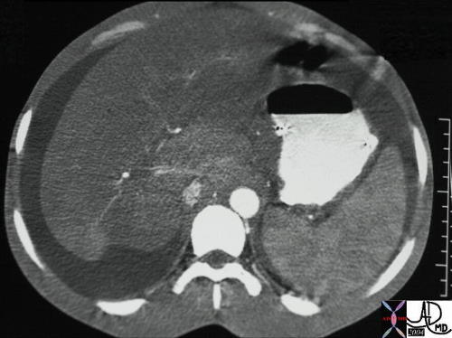
Budd-Chiari syndrome is an acute, chronic or subacute thrombotic or obstructive circulatory disorder of the hepatic venous system cause multiple factors including hypercoaguable states masses around the hepatic veins resulting in thrombosis of the hepatic veins sinusoids or IVC characterised by liver congestion divided into ivc origin hepatic venous origin sinusoidal origin pathogenesis disorder causes liver injury results in variable outcome resolution fibrosis acute or chronic liver failure structural disorder chronic congestion functional disorder variable depending on ability to develop collateral clinical presentation pain ascites diagnostic studies include MRI US, CT, angiography treatment is commonly by anticoagulation
| 25225 |
| 25225 Courtesy Ashley Davidoff MD code liver + caudate lobe enlarged normal perfusion collaterals fx heterogeneous + dx Budd Chiari syndrome + imaging radiology CTscan C+ |
| 25231 |
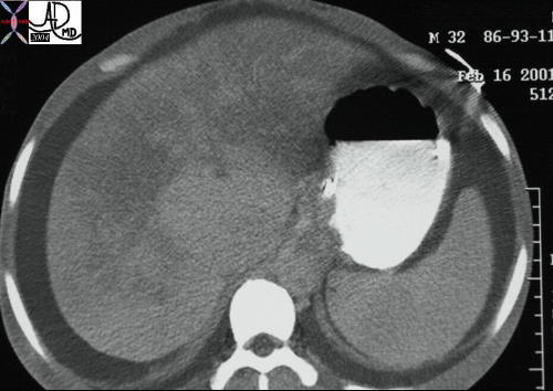
| 25231 liver + caudate lobe + fx large + fx isolated enhancement + dx Budd Chiari syndrome + imaging radiology CTscan C+ |
| 25243 |
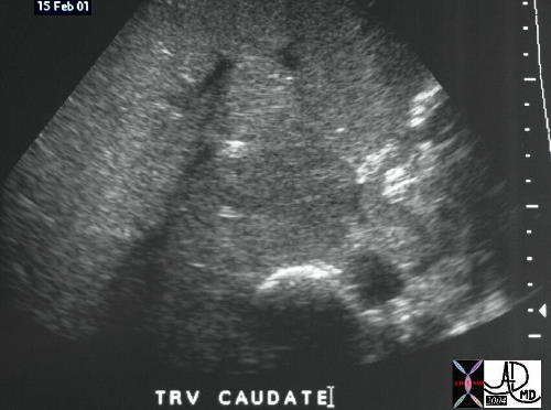
| 25243 liver + caudate lobe + fx large + dx Budd Chiari syndrome + imaging radiology USscan |
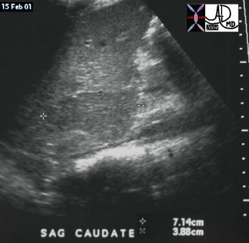
Budd Chiari syndrome |
| 25245 liver + caudate lobe + fx large + dx Budd Chiari syndrome + imaging radiology USscan |

Tricuspid Regurgitation into the Hepatic Veins |
| 48098 heart cardiac liver enlarged hepatic veins fx reflux into the hepatic veins dx tricuspid regurgitation tricuspid valve congestive heart failure CHF CTscan Davidoff MD 48098c01 48098c02 48089b01 |
|