Atherosclerosis
Ashley Davidoff MD
The Common Vein Copyright 2012
Definition
Atherosclerosis is a degenerative disorder of the intima of arteries. (occasionally affecting other structures in the cardiovascular system) chatracterised by accumulation of fatty and fibrous elements within the intima of the vessel wall. The multifactorial causes of atherosclerosis include age, sex, genetics, lifestyle and dietary factors. among many other unfolding factors. The recognizable “phases” of atherosclerosis include ; 1) breach of the endothelium (33792d) 2) migration of lipoproteins from the lumen into the intima. (33792d) 3) Formation of a lipoprotein-proteoglycan complex that traps the lipoprotein in the intima (33792e) 4) Migration of leukocytes from the lumen into the intima. (33792g) 5) Transformation of the monocytes into macrophages and phagocytosis of the fat complexes to form fat laden foamy macrophages. (33792h) 6) Migration of smooth muscle cells from the media into the intima and transformation into fibrocytes. A fibrous capsule around the fatty complex is formed. (33792i, 33801b) 7) Cell death and destruction with associated formation of dystrophic calcification. 8) Growth of the atheromatous complex with impingement on the lumen. (33801b) 9) Potential of the complex to rupture. (33801d) The progressive impingement on the lumen causes reduction of blood flow, usually manifesting clinically with angina when the lumen is reduced by 70% of its original size. Acute rupture of a plaque predisposes to superadded thrombosis and occlusion of the lumen. Acute myocardial infarction and death are complications of this event. The initial phase of endothelial injury has multifactorial contributing factors including hypertension, hemodynamic factors, hyperlipidemia, homocysteine, smoking, toxins, immune reactions, and viral disease. The result is a breach in the intima and migration of lipoproteins into the subendothelial layer. The lipoproteins and proteoglycans form a complex compound that traps the fat complex in the subepithelial layer. The next phase heralds the migration of leukocytes and platelets into the subendothelial layer. The monocytes, phagocytose the lipoprotein-proteoglycan complex forming the lipid laden foamy histiocytes. These macrophages can apparently repllicate. At this stage the macroscopic appearance is the well known “fatty streak”. The next phase of atherosclerosis occurs following smooth muscle proliferation and migration from the media into the evolving plaque. Smooth muscle also undergoes apoptosis and death. The vascular smooth muscle produces the extracellular matrix has major contribution to the atheromatous lesion. Interstitial collagens (types I and III) and proteoglycans such and elastin fibers also accumulate in atherosclerotic plaques. Accumlation of cholesterol and cholesterol esters occur. Smooth muscle migrates from the media into the intima and transform into fibrous elements both within the lesion as well superficial to the lesion forming a fibrous plaque.The monocytes and macrophages ingest the fat and foamy fat laden macrophages result. An inflammatory reaction evolves. Continued accumulation of lipid, macrophages, foamy cells, extracellular matrix result in a fibrofatty complex that grows in size over time. As it enlarges it bulges and thins the intima and eventually erodes the surface. As the disease progresses, cell death within the plaque results, and dystrophic calcification follows.
Background
 Normal Lipoproteins in the Circulation Normal Lipoproteins in the Circulation |
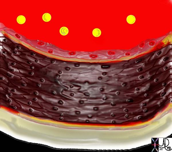
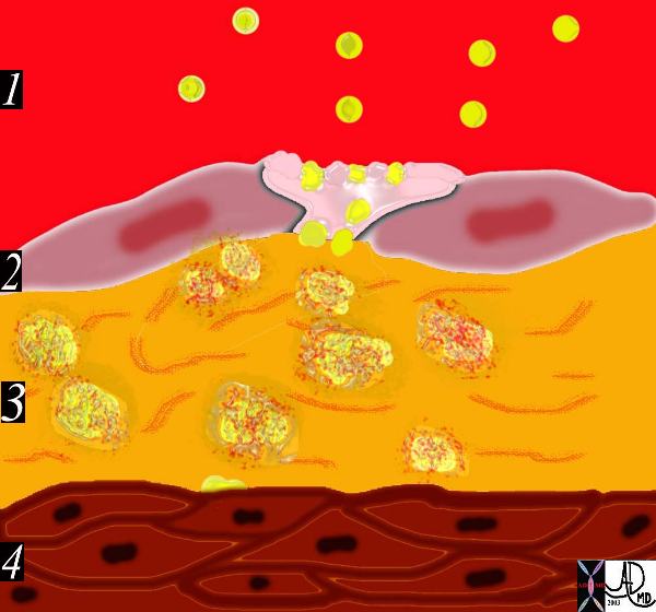
| This image shows normal circulating lipoproteins in the circulation. The intima is intact and the wall is normal.he factors the presence of lipoprotein molecules in the lumen.
Copyright 2012 Courtesy Ashley Davidoff MD. 33789 Davidoff art |
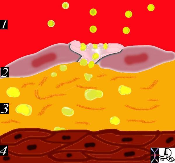
The circulating lipoproteins enter a breached endothelium (2) and enter the subendothelial layer of supporting connective tissue within which are linear starands of proteoglycan. (3). At this stage the media (4) is quiescent. Courtesy Ashley Davidoff MD 33792d code heart artery intima endothelium histopathology pathogenesis atherosclerosis atheroma drawing
atheroma drawing
 The Aging Artery The Aging Artery
An Observation by da Vinci |
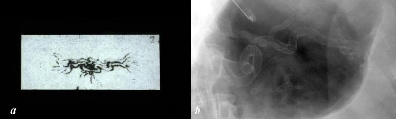
| The drawing of the “meseraic vessels” – presumably the celiac axis is by Leonardo da Vinci. Da Vinci was able to interview and perform an autopsy on a centenarian. He describes the desiccated and tortuous state of the vessels of this patient accurately depicting the atherosclerotic process. He describes the narrowed lumen and the consequences of poor blood flow. The translation of da Vinci’s text accompanying this one inch image is remarkably insightful and pioneering. (see atherosclerosis historical da Vinci) Image b is a magnified view of the left upper quadrant showing a calcified serpiginous splenic artery much like the atherosclerotic vessel described by da Vinci.
Courtesy Ashley Davidoff MD 13045b da Vinci diagram 113318c.8L |
This diagram shows the yellow spheroidal lipoproteins traverse the injured epithelium (2) from the lumen (red) and binding to the linear shaped proteoglycan molecules in the intimal layer. (3) In essence the lipoprotein is “captured”, because it has been altered structurally and is unable to return to the circulation. Courtesy Ashley Davidoff MD. 33792e code heart artery intima endothelium histopathology pathogenesis atherosclerosis atheroma drawing
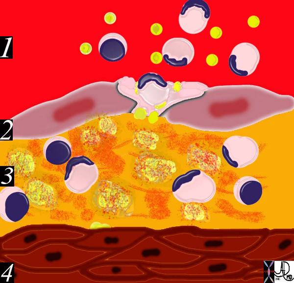 The diagram shows an evolving atheromatous plaque with inflammatory cells, monocytes, and lymphocytes infiltrating the intima which contains the lipoprotein- proteoglycan complex, extracellular lipid , and cholesterol crystals. 33792g Courtesy Ashley Davidoff MD. code heart artery intima endothelium histopathology pathogenesis atherosclerosis atheroma drawing
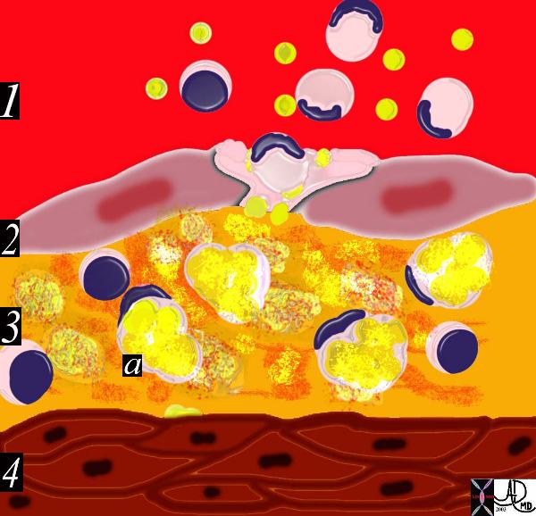 This drawing shows migration of the monocytes into the intima. These monocytes transform into macrophages in the intima and phagocytose the lipid products to become lipid laden foamy cells. The macroscopic correlate at this stage is the fatty streak. Courtesy Ashley Davidoff MD 33792h . code heart artery intima endothelium histopathology pathogenesis atherosclerosis atheroma drawing
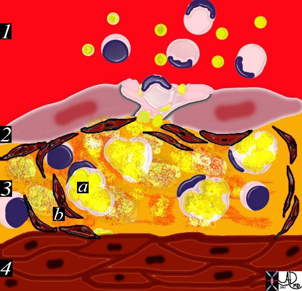
This diagram shows the reaction of the smooth muscle cells (b) to the formation of foam cells (a) in the subendothelial layer of the intima. The smooth muscle cells migrate from the muscular layer (4) into the intima. Here they undergo dedifferentiation into fibrocytes. 33792i Courtesy Ashley Davidoff MD. code heart artery intima endothelium histopathology pathogenesis atherosclerosis atheroma drawing
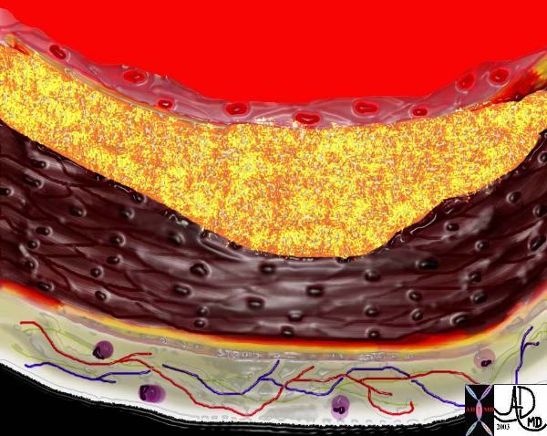
The diagram shows the atherosclerotic lesion in the subepithelial layer of the intima which at first bulges toward the media or muscular layer. 33801a Courtesy Ashley Davidoff MD. code heart artery intima endothelium histopathology pathogenesis atherosclerosis atheroma drawing
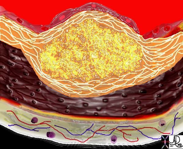
The diagram shows the atherosclerotic lesion in the subepithelial layer of the intima which is bulging both toward the media and toward the lumen. There is a central core of fat and necrotic debris, surrounded by fibrous elements which give the plaque its hardness to the feel. The accumulation of fibrous tissue heralds an advanced atherosclerotic lesion. 33801b Courtesy Ashley Davidoff MD. code heart artery atherosclerosis
Historical
“da Vinci had the unique experience of being able to know and perform an autopsy on an centenarian and described his findings of the aorta and splenic artery – characteristic of atherosclerosis. He observed the tortuosisy of the vessels, the calcification, the aneurysmal nature, the hardening and the lumenal narrowing of the atherosclerotic process. The following is the direct translation of daVinci’s writing. “The artery and vein which in the aged, extend between the spleen and the liver, acquire so thick a covering that it contracts the passage of the blood which comes from the meseraic (mesenteric portal) veins. By means of these veins the blood through the liver to the heartand the two major veins (cava) and consequently through the entire body. These veins apart from the thickening grow in length and twist like a snake, ….. …I have found there stones in the vessels which pass beneath the clavicles of the chest. These were as large as chestnuts of the color and shape of truffles or of dross or clinkers of iron. These stones were extremely hard, like these clinkers, and had formed sacs attached to the said vessels, in the manner of goiters. …And this old man, a few hours before his death, told me that he had passed one hundred years, and that he was concious of no failure of body, except feebleness. And thus sitting upon a bed in the hospital of Santa Maria Nuova Florence, without any untoward movement or sign, he passed from this life. And I made an anatomy to see the cause of death to see the cause of death so sweet, which I found to proceed from debility through lack of blood and deficiency of the artery (aorta) which nourishes the heart and other lower members. I found this artery very dessicated, shrunken and withered. This anatomy I described very carefully, and with great ease owing to the absence of fat and humor which rather hinder recognition of the parts. The other anatomy was that of a child of 2 yeqrs in which I found everything to be oppposite to that old man. The aged who enjoy good health die through lack of nourishment. This happens because the lumen of the meseraic (mesenteric portal) veins is constantly constricted by the thickening of the coverings of these vessels; which progress as far as the capillary vessels (vene chapillari) which are the first to close up entirely. As a consequence of this, the old dread the cold more than the young and those who are extremely old have a skin the color of wood or of dry chestnut because the skin is almost completely deprived of nourishment. The tunics of the vessels behave in man as in oranges, in which the peel thickens and the pulp diminishes the older they become. And if you say that it is the thickened blood which does not flowthrough the vessels, this is not true, for the blood does not thicken in the vessels because it is continually dies and is renewed.”