Hemorrhage
Copyright 2009
Definition
Hemorrhage is …
characterized by …..
caused by ..etiology or predisposing factors
resulting in a pathological feature (structural change or functional change) or clinical feauture
Sometimes complicated by ….
Diagnosis is suspected clinically by … and confirmed by ….
Imaging includes the use of
Treatment options depend on …. but includes …..
Etymology if available
Principles
Hemorrhage is escape of blood from blood vessels.
Size Shape Position Character Time Space Diseases
 
Intact River and Rupture and Flooding |
| 83567.800 Charles river Boston Massachusetts Salt and Pepper Bridge order TCV The Common VEin Davidoff photography
|
Size
Small Contusion
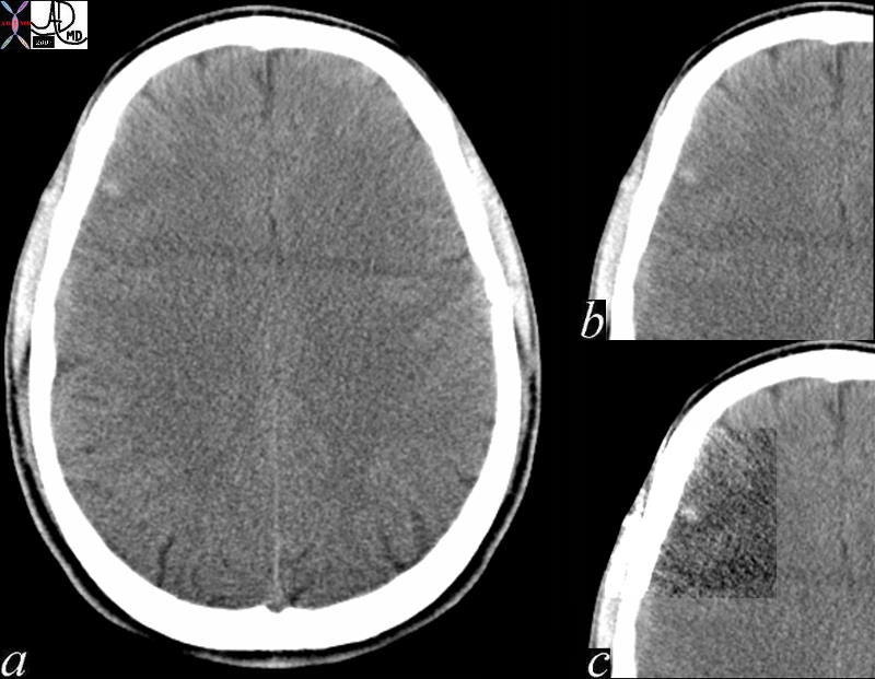
Brain Contusion |
| 70048c01 brain parietal lobe fx hyperdensity small halo edema dx traumatic cerebral contusion bruise injury of brain without a break in the skin. L. Contusio, from contundere = to bruise CTscan 70048c01 70048.800 Davidoff MD |
Shape
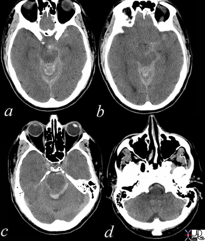
Subarachnoid Bleed – Ruptured Aneurysm |
| 72034c01 59 male presents with headache brain meninges tentorium circle of Willis ambient cisterns Berry aneurysm rupture subarachnoid blood subarachnoid hemorrhage headache CTscan Davidoff MD |
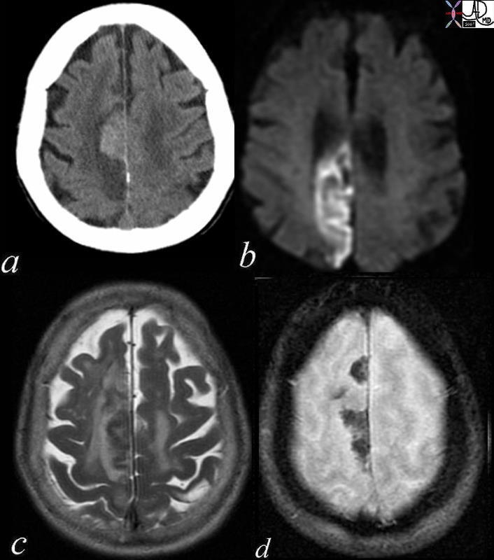
Acute Hemorrhagic Infarction |
| 71239c01 patient with atrial fibrillation brain cerebrum parietal lobe increased density hyperdense parasagittal hemorrhagic infarct paracentral DWII bright T2 bright GRE mixed blood products dx acute hemorrhagic infarction secondary to embolic event the patient disd have other non hemorrhagic infrctions in other parts of the brain CTscan MRI Davidoff MD |
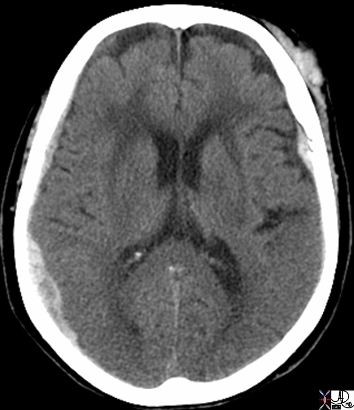
Bilateral Subdural Hematomas |
| 70000.800 skin brain cerebrum cerebral subcutaneous subdural contra coup fx acute blood fx density dense subdural hematoma subcutaneous hematoma trauma radiologists and detectives CTscan Davidoff MD |
Position
CNS

Subarachnoid Bleed – Ruptured Aneurysm |
| 72034c01 59 male presents with headache brain meninges tentorium circle of Willis ambient cisterns Berry aneurysm rupture subarachnoid blood subarachnoid hemorrhage headache CTscan Davidoff MD |
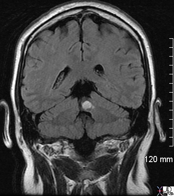
Acute Hemorrhage in the Left Cerebellar Vermis |
| 71730 male with acute ataxia roof of fourth 4th ventricle acute bleed left vermis venous angioma brain cerebellum vermis vein cerebellar venous malformation vascular malformation T1 pre gadolinium T1 bright lesion dx acute hemorrhage MRI Davidoff MD |
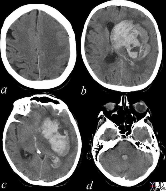
Acute Cerebral Hemorrhage with Mass Effect and Rupture into the Ventricles |
| 72143c01 brain cerebral hemorrhage hemorrhagic CVA hematoma lateral ventricles 4th ventricle fourth ventricle mass effect gray matter white matter loss of the gray white interface cerebral edema blood CTscan CVA Davidoff MD |
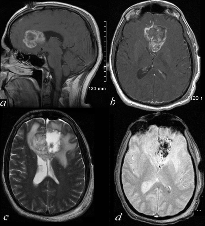
Glioblastoma of the Corpus Callosum S/P Debulking |
| 72420c01 46 male brain cerebrum genu of the corpus callosum crosses midline enhancing tumor in periphery surrounding edema dilated right hor mass effect on the lateral ventricles tumor arises from the corpus callosum post operative resection of the left side of the tumor methemoglobin effects in the left side a = T1 post gadolinium b= T1 weighted post contrast c= T2 weighted d = GRE blood products T2* effects dx glioblastoma s/p debulking Davidoff MD |
Character
CVS
Inflammation
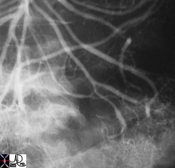 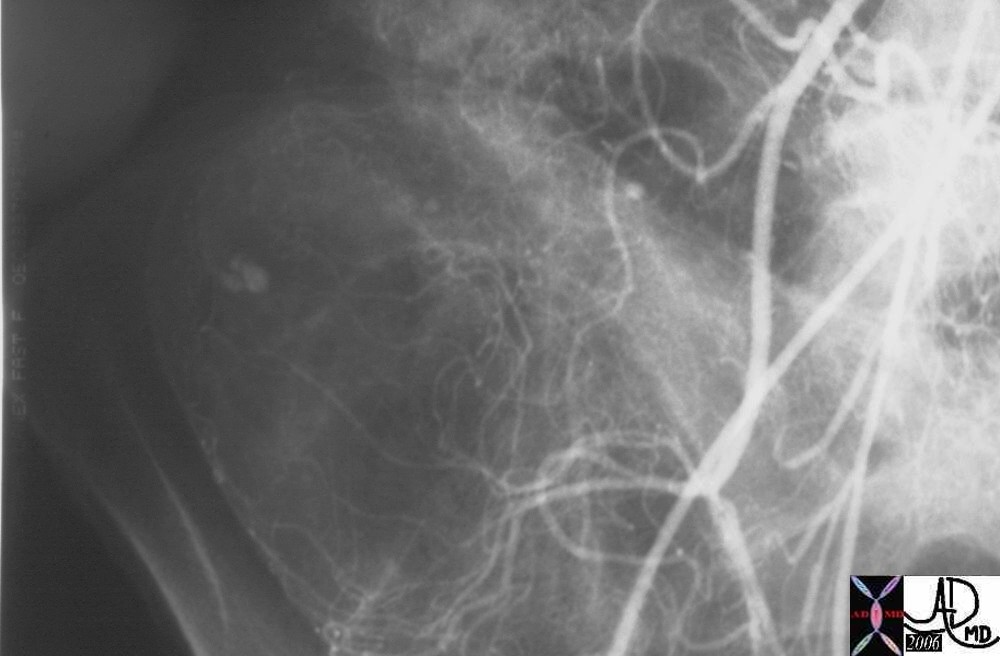
Henoch Schonlein Arteritis Segmental Spasm with Hemorrhage |
| Young female presents with GI bleed hemorrhage bleeding blood small bowel colon SMA superior mesenteric artery jejunal branches ileocolic artery fx arterial spasm fx contrast extravasation RLQ in cecum dx arteritis angiitis vasculitis arteriopathy dx Henoch -Schonlein arteritis angiography angiogram Courtesy Ashley Davidoff MD 28514 28515 28516 28517 surgical specimen showed plaque like ulcers in the bowel consistent with chronic ischemia |
Mechanical Disorders
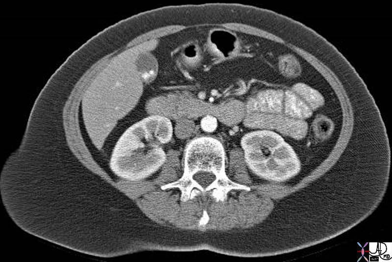 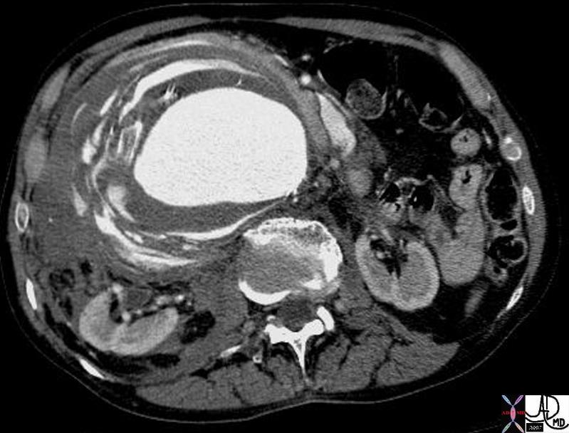
Normal and Ruptured Aorta |
| 37004 abdomen aorta kidneys cortical phase subcurtaneous fat adipose tissue normal anatomy gallsones cholelthiasis CT scan Davidoff MD
18269 aorta abdomen AAA aortic aneurysm abdominal aorta retroperitoneum fx retroperitoneal hematoma fx active hemorrhage fx perinephric hematoma anterior pararenal space perirenal space posterior pararenal space hemorrhage dx rupture abdominal aortic aneurysm CTscan Davidoff MD fx ruptured AAA |
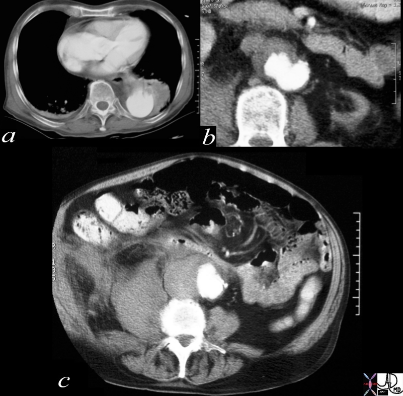
Penetrating Ulcer with Rupture |
| 17529c01 artery descending thoracic aorta abdominal aorta dx rupture pseudoaneumysm ulcerating plaque mural hematoma ruptured through aortic wall hemorrhage hematoma retroperitoneum CTscan Courtesy Ashley DAvidoff MD Ashley Davidoff MD |
RS
GIT
Inflammation
|
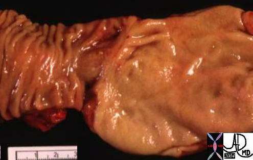
Normal Antrum and Stomach with Hemorrhagic Gastritis |
| 01978b stomach gastric pylorus duodenum junction normal anatomy
12263 code stomach gastric + hemorrhagic gastritis grosspathology |
|
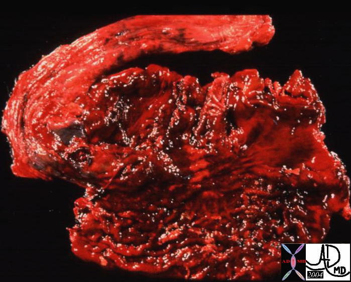
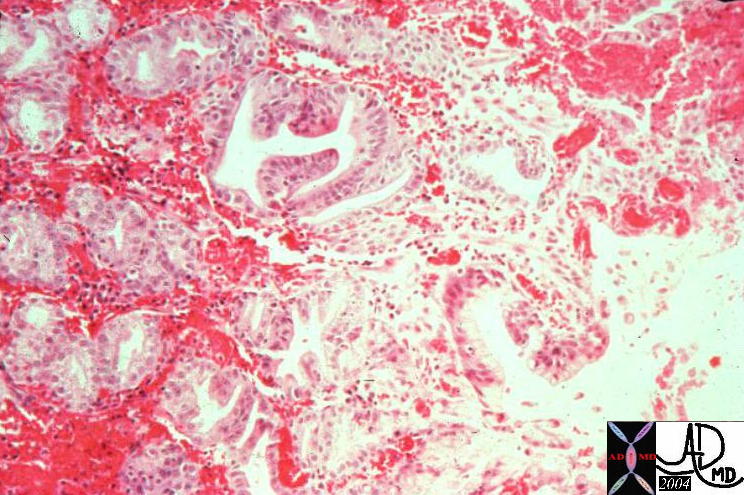
Hemorrhagic Gastritis |
| 12264 code stomach gastric + hemorrhagic gastritis grosspathology |
GUT
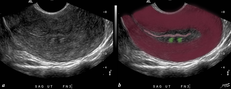
Two Lesions in the Junctional Zone Impinging on the Endometrium |
| This patient presents with menorrhagia. Two echogenic nodules (overlaid in green) are seen in the subendometrial layer, (junctional zone). They are in close proximity and do appear to have appositional relationships with the endometrial stripe. They appear to and distort the endometrial lining. These findings could account for the patient’s menorrhagia. Note that the uterus is retroverted Included in the differential diagnosis are submucosal fibroids, and dystrophic changes in prior foci of adenomyosis. An MRI would be helpful in further characterizing these lesions in the subendometrial layer
copyright 2009 all rights resrved Courtesy Ashley Davidoff MD 85641bc01.8s |
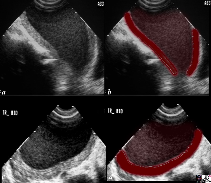
Hemorrhage into the Endometrial Cavity |
| The ultrasound is from a 33 year old female who had a cervical biopsy and then showed complex fluid in a distended endometrial cavity. the findings are consistent with hematocolpos, but in the appropriate clinical setting could represent pyocolpos
uterus blood hematocoplos expanded cavity complex fluid Courtesy Ashley DAvidoff MD copyright 2009 all rights reserved 85940c02.8s |
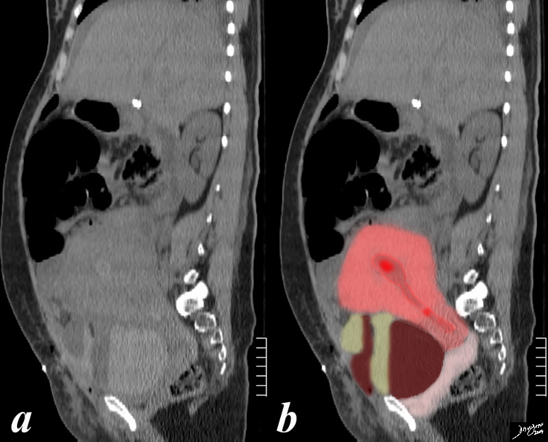
Post Cesarian Section Hemorrhage in Endometrial Cavity Hemorrhage into Pelvic Cavity |
| The CTscan is from a 28 year old female following a cesarian section showing a small amount of blood in the endometrial cavity and two large collections anterior to the low uterine segment. It is likely that the blood is from the incision of the LUS. A Hematocrit level is characterized by a clot (maroon)/fluid (serum yellow) level. Free fluid anterior to the liver is also noted.
uterus enlarged post partum endometrium hemorrhage fluid fluid level peritoneal cavity intraperitoneal blood retained products considered as well cesarian section CTscan Courtesy Ashley Davidoff MD copyright 2009 all rights reserved 84070c02.81s |
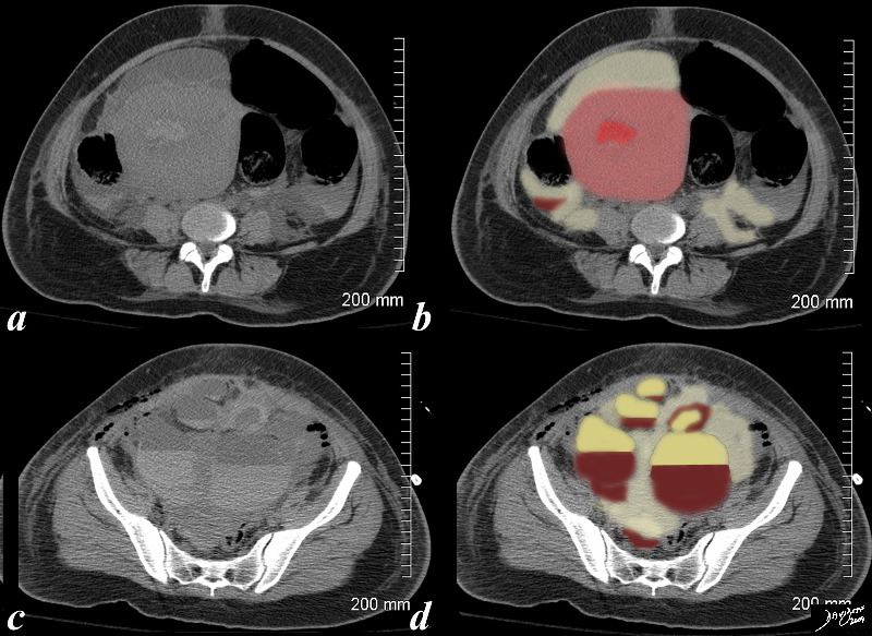
Post Cesarian Section Hemorrhage in Endometrial Cavity Hemorrhage into Pelvic Cavity |
| 84070c.8s The CTscan is from a 28 year old female following a cesarian section showing a small amount of blood in the endometrial cavity and multiple large collections in the pelvist. It is likely that the blood is from the incision of the LUS. Hematocrit levels are characterized by sedimented red cells (maroon)/fluid (serum yellow) level. Free fluid (lighter yellow) is also noted.
uterus enlarged post partum endometrium hemorrhage fluid fluid level peritoneal cavity intraperitoneal blood retained products considered as well cesarian section CTscan Courtesy Ashley Davidoff MD copyright 2009 all rights reserved 84070c02.81s |
MS
Skin

Bilateral Subdural Hematomas |
| 70000.800 skin brain cerebrum cerebral subcutaneous subdural contra coup fx acute blood fx density dense subdural hematoma subcutaneous hematoma trauma radiologists and detectives CTscan Davidoff MD |
RES
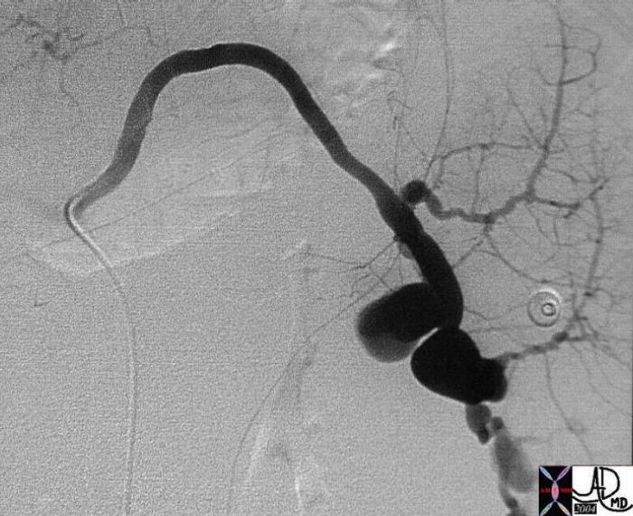 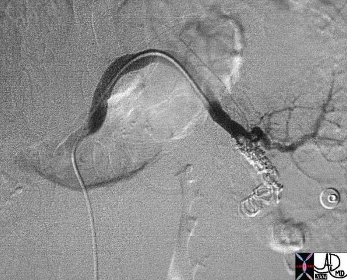
Ruptured Aneurysm and Repair by Embolization |
| 22521 22524 Courtesy Ashley Davidoff MD code spleen + artery + dx ruptured splenic artery aneurysm pseudoaneurysm + cystic fibrosis biliary cirrhosis portal hypertension angiography emergency |
Character
Space
Time
 
Normal and Ruptured Aorta |
| 37004 abdomen aorta kidneys cortical phase subcurtaneous fat adipose tissue normal anatomy gallsones cholelthiasis CT scan Davidoff MD
18269 aorta abdomen AAA aortic aneurysm abdominal aorta retroperitoneum fx retroperitoneal hematoma fx active hemorrhage fx perinephric hematoma anterior pararenal space perirenal space posterior pararenal space hemorrhage dx rupture abdominal aortic aneurysm CTscan Davidoff MD fx ruptured AAA |
 2 2
Left Sided Ruptured Hemorrhagic Cyst with Free Blood in the Pelvis and Bleeding onto the Greater Omentum RLQ (pink) |
| This CT scan is that of a 27 year old female who presented with acute lower pelvic pain in mid cycle. The findings of free blood in the pelvidss (maroon) the cyst (yelloow) with an enhancing rim9Bright red) and spillage of b;lood onto the greater omentum (pink let anterior) are consistent with a ruptured hemorrhagic cyst .
24480c01 27 year old female presented with lower abdominal pain pelvic pain ovary fx cyst cul de sac blood free blood hyperdense corpus luteum cyst greater ometum congested fx enhancing dx hemorhagic ovarian cyst CT scan C- CTscan Courtesy Ashley DAvidoff MD |
Diseases
Trauma
Thrombus – Acute

Bilateral Subdural Hematomas |
| 70000.800 skin brain cerebrum cerebral subcutaneous subdural contra coup fx acute blood fx density dense subdural hematoma subcutaneous hematoma trauma radiologists and detectives CTscan Davidoff MD |
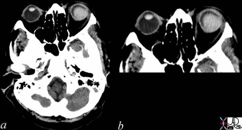
Eye – Hemorrhage |
| 72040c01 eye globe fx dense conjunctiva fx thickened fx hyperdense dx acute hemorrhage blood bleed CTscan Davidoff MD |
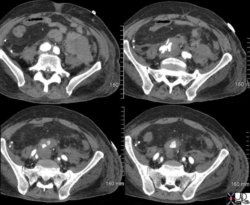
Ruptured Pseudoaneurysm of the Right Common Iliac Artery |
| 75727c01 elderly man with acute pelvic and back pain aorta iliac artery RLQ right lower quadrant artery pseudoaneurysm ruptured aneurysm retroperitoneal hematoma hemorrhage CTscan Courtesy Ashley Davidoff MD Courtesy Rebecca Schwartz MD 75727c01 75727c02 75724c01 75727c03 |
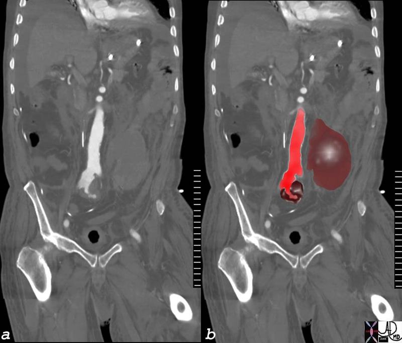
Ruptured Pseudoaneurysm of the Right Common Iliac Artery |
| 75724c01 elderly man with acute pelvic and back pain aorta iliac artery RLQ right lower quadrant artery pseudoaneurysm ruptured aneurysm retroperitoneal hematoma hemorrhage CTscan Courtesy Ashley Davidoff MD Courtesy Rebecca Schwartz MD 75727c01 75727c02 75724c01 75727c03 |
Time
Intracranial
CT
(change from HYPER- to HYPO- dense over time)
Initially – 60 – 90 Hounsfield Units (HU)
2 days – 70 HU
3 weeks – 30 HU
>5 weeks – <30 HU
20% show enhancing rim at 2-6 weeks
Reference Medpix USUHS
MRI
(T1/T2: II, ID, BD, BB, DD (I-iso, D-dark, B-bright)
Hyperacute – minutes to hours (DD => II)
– T1WI – hematoma hypointense (deoxyHb) => isointense
– T2WI – hematoma hypointense (deoxyHb) => isointense
Acute – 0-2 days (ID => BD)
– deoxyhemoglobin in intact RBCs with surrounding edema
– T1WI – hematoma isointense, low signal intensity (SI) edema
– T2WI – hematoma decreased SI at center, high SI edema
Subacute – 2-14 days (BB)
– deoxyhemoglobin changes to methemoglobin from outer to inner
– T1WI – outer core shows increased SI
– T2WI – Outer core shows increased SI due to shortened T1, longer T2
Chronic – 14 days (BB => DD)
– hemosiderin laden macrophages at periphery
– T1WI – inner core now also increased SI, rim has low SI
– T2WI – inner core also has increased SI, rim has low SI
Chronic – months later (DD)
– hemosiderin laden macrophages at periphery
– T1WI – mostly iso-/decreased SI, rim has lower SI
– T2WI – markedly hypointense rim has low SI – “blooms” with greater T2-weighting
Reference Medpix USUHS

