The Common Vein Copyright 2010
Definition
|
Acute Multicentric Non Hemorrhagic Infarcts Consistent with Embolic Disease |
|
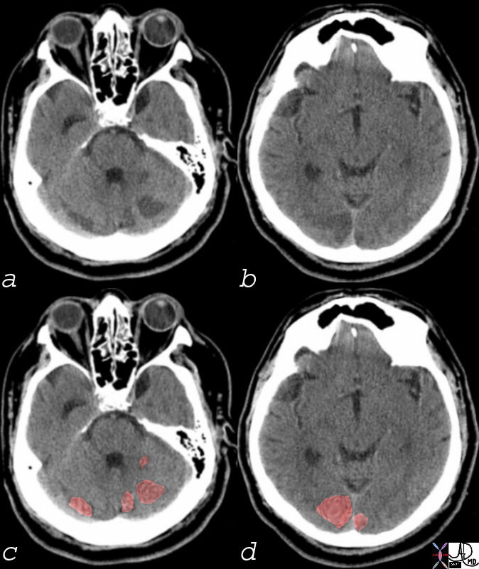
Multicentric Infarcts in the Cerebellum and Occipital Lobes Multicentric low density regions in both posterior cerebellar hemispheres and the occipital lobes bilaterally seen in (a and b) correspondingly overlaid in light red in c and d, are consistent with non hemorrhagic infarcts. Embolic disease is most likely Courtesy Ashley Davidoff MD 74923c01 |
Non Hemorrhagic Emboli

Carotid Stenosis and Acute Multicentric Non Hemorrhagic Emboli |
|
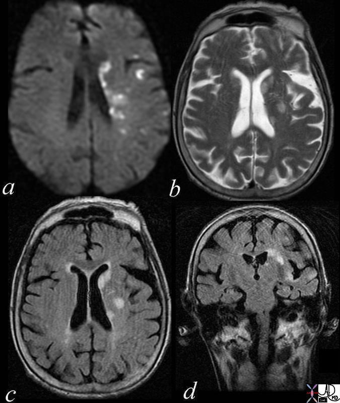
Multicentric Infarcts – Carotid Stenosis The images reveal multicentric acute infarcts in the left cerebral hemisphere involving the internal capsule and left parietal lobe cortex. Image a is the DWI sequence and the high intensity foci are diagnostic of acute infarction. Image b is a T2 weighted image that reflects increase water in the high intensity regions. Image c is an axial FLAIR and d a coronal FLAIR sequence both sensitive to the regions of infarct and characerized by high intensity foci in the regions of acute infarction. Courtesy Ashley Davidoff MD 72014c01 |
Hemorrhagic Manifiestations of Cerebral Emboli

Atrial Fibrillation and Hemorrhagic Emboli |
|
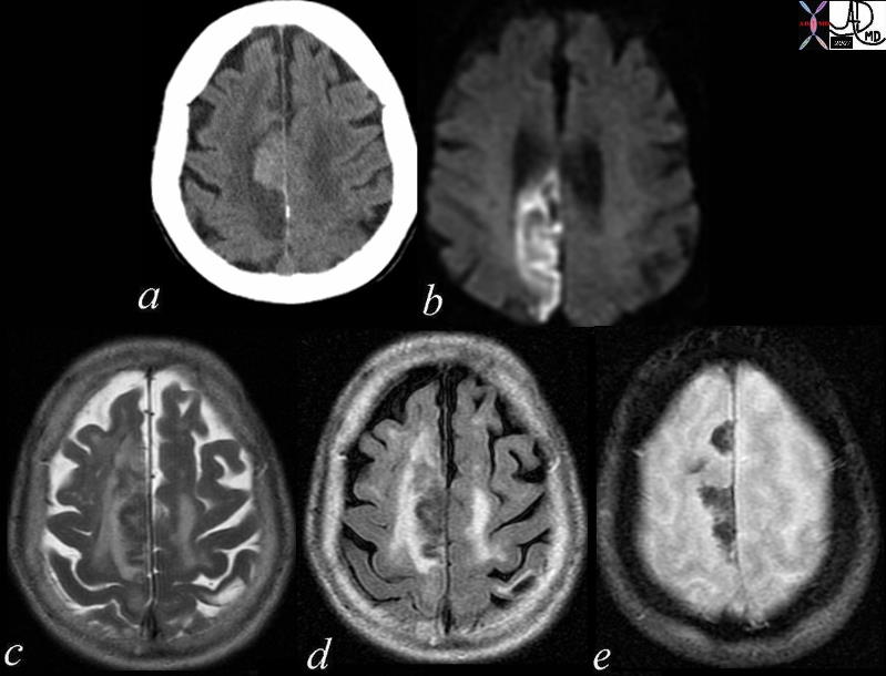
The axial images are from a patient with atrial fibrillation and neurological deficits. Image a is a CT scan which shows a high density lesion in the vertex of the right parietal lobe suggesting hemorrhagic change. Image b is a diffusion weighted MRI image at the level of the ventricles which shows a high intensity region in the parieto-occipital region suggesting acute infarction. Image c is a axial T2 weighted image showing edema in the white matter of the right parietal lobe. Image e is a GRE image showing mixed heterogeneity with probable iron deposition suggesting subacute or chronic hemorhage. Findings are consistent with old and new multicentric infarcts of the brain likely from the heart caused by atrial fibrillation Courtesy Ashley Davidoff MD 71239c02 |

Acute Hemorrhagic Infarct – Multicentric – Likely Embolic |
|
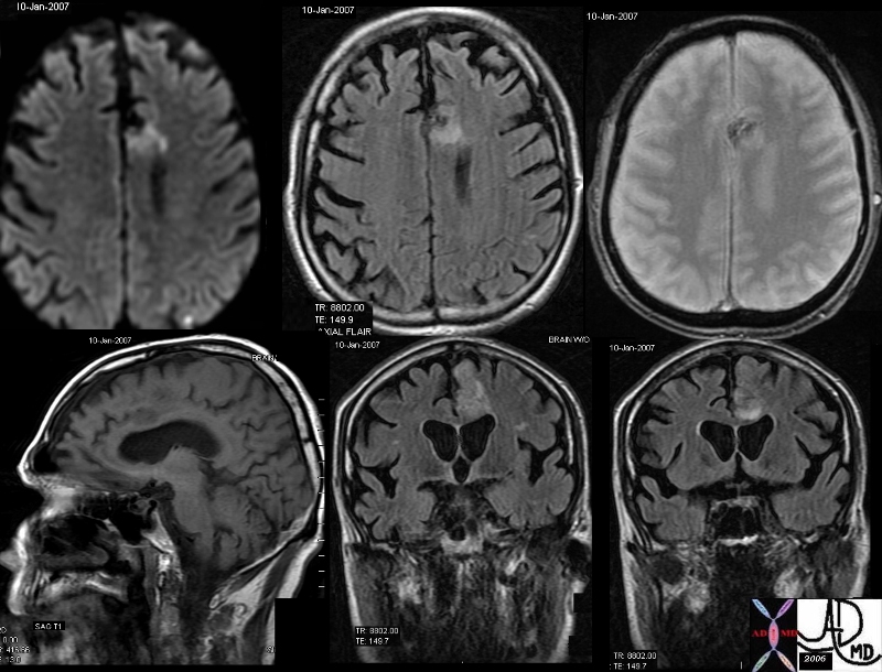
The MRI in various projections confirms the presence of an acute hemorrhagic infarct in the left paracentral frontal territory. The diffusion images (top left), show a hyperintense focus in the left frontal lobe, which is hyperintense on the axial FLAIR image (middle top), hypointense and granular on the GRE (top right), hypointense on the sagittal T1 (bottom left) and seen as hyperintense on the last two STIR coronal images. These findings are consistent with an acute hemorrhagic focus which and was thought to be embolic in origin due to other noted foci. Courtesy Ashley Davidoff MD copyright 2010 46098c01.800 |

